MRI
-
 KAIST Develops Analog Memristive Synapses for Neuromorphic Chips
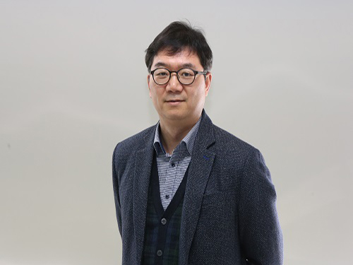
(Professor Sung-Yool Choi from the School of Electrical Engineering)
A KAIST research team developed a technology that makes a transition of the operation mode of flexible memristors to synaptic analog switching by reducing the size of the formed filament. Through this technology, memristors can extend their role to memristive synapses for neuromorphic chips, which will lead to developing soft neuromorphic intelligent systems.
Brain-inspired neuromorphic chips have been gaining a great deal of attention for reducing the power consumption and integrating data processing, compared to conventional semiconductor chips. Similarly, memristors are known to be the most suitable candidate for making a crossbar array which is the most efficient architecture for realizing hardware-based artificial neural network (ANN) inside a neuromorphic chip.
A hardware-based ANN consists of a neuron circuit and synapse elements, the connecting pieces. In the neuromorphic system, the synaptic weight, which represents the connection strength between neurons, should be stored and updated as the type of analog data at each synapse.
However, most memristors have digital characteristics suitable for nonvolatile memory. These characteristics put a limitation on the analog operation of the memristors, which makes it difficult to apply them to synaptic devices.
Professor Sung-Yool Choi from the School of Electrical Engineering and his team fabricated a flexible polymer memristor on a plastic substrate, and found that changing the size of the conductive metal filaments formed inside the device on the scale of metal atoms can make a transition of the memristor behavior from digital to analog.
Using this phenomenon, the team developed flexible memristor-based electronic synapses, which can continuously and linearly update synaptic weight, and operate under mechanical deformations such as bending.
The team confirmed that the ANN based on these memristor synapses can effectively classify person’s facial images even when they were damaged. This research demonstrated the possibility of a neuromorphic chip that can efficiently recognize faces, numbers, and objects.
Professor Choi said, “We found the principles underlying the transition from digital to analog operation of the memristors. I believe that this research paves the way for applying various memristors to either digital memory or electronic synapses, and will accelerate the development of a high-performing neuromorphic chip.”
In a joint research project with Professor Sung Gap Im (KAIST) and Professor V. P. Dravid (Northwestern University), this study was led by Dr. Byung Chul Jang (Samsung Electronics), Dr. Sungkyu Kim (Northwestern University) and Dr. Sang Yoon Yang (KAIST), and was published online in Nano Letters (10.1021/acs.nanolett.8b04023) on January 4, 2019.
Figure 1. a) Schematic illustration of a flexible pV3D3 memristor-based electronic synapse array. b) Cross-sectional TEM image of the flexible pV3D3 memristor
2019.02.28 View 9228
 -
 A Parallel MRI Method Accelerating Imaging Time Proposed
KAIST researchers proposed new technology that reduces MRI (magnetic resonance imaging) acquisition time to less than a sixth of the conventional method. They made a reconstruction method using machine learning of multilayer perception (MLP) algorithm to accelerate imaging time.
High-quality image can be reconstructed from subsampled data using the proposed method. This method can be further applied to various k-space subsampling patterns in a phase encoding direction, and its processing can be performed in real time.
The research, led by Professor Hyun Wook Park from the Department of Electrical Engineering, was described in Medical Physics as the cover paper last December. Ph.D. candidate Kinam Kwon is the first author.
MRI is an imaging technique that allows various contrasts of soft tissues without using radioactivity. Since MRI could image not only anatomical structures, but also functional and physiological features, it is widely used in medical diagnoses. However, one of the major shortcomings of MRI is its long imaging time. It induces patients’ discomfort, which is closely related to voluntary and involuntary motions, thereby deteriorating the quality of the MR images. In addition, lengthy imaging times limit the system’s throughput, which results in the long waiting times of patients as well as the increased medical expenses.
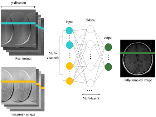
To reconstruct MR images from subsampled data, the team applied the MLP to reduce aliasing artifacts generated by subsampling in k-space. The MLP is learned from training data to map aliased input images into desired alias-free images. The input of the MLP is all voxels in the aliased lines of multichannel real and imaginary images from the subsampled k-space data, and the desired output is all voxels in the corresponding alias-free line of the root-sum-of-squares of multichannel images from fully sampled k-space data. Aliasing artifacts in an image reconstructed from subsampled data were reduced by line-by-line processing of the learned MLP architecture.
Reconstructed images from the proposed method are better than those from compared methods in terms of normalized root-mean-square error. The proposed method can be applied to image reconstruction for any k-space subsampling patterns in a phase encoding direction. Moreover, to further reduce the reconstruction time, it is easily implemented by parallel processing.
To address the aliasing artifact phenomenon, the team employed a parallel imaging technique using several receiver coils of various sensitivities and a compressed sensing technique using sparsity of signals.
Existing methods are heavily affected by sub-sampling patterns, but the team’s technique is applicable for various sub-sampling patterns, resulting in superior reconstructed images compared to existing methods, as well as allowing real-time reconstruction.
Professor Park said, "MRIs have become essential equipment in clinical diagnosis. However, the time consumption and the cost led to many inconveniences." He continued, "This method using machine learning could greatly improve the patients’ satisfaction with medical service." This research was funded by the Ministry of Science and ICT.
(Firgure 1. Cover of Medical Physics for December 2017)
(Figure 2. Concept map for the suggested network)
(Figure 3. Concept map for conventional MRI image acquisition and accelerated image acquisiton)
2018.01.16 View 6493
 -
 The key to Alzheimer disease, PET-MRI made in Korea
Professor Kyu-Sung Cho
- Simultaneous PET-MRI imaging system commercialization technology developed purely from domestic technology - - Inspiring achievement by KAIST, National NanoFab Center, Sogang University, Seoul National University Hospital –
Hopes are high for the potential of producing domestic products in the field of state-of-the-art medical imaging equipment that used to rely on imported products.
The joint research team (KAIST, Sogang University and Seoul National University) with KAIST Department of Nuclear and Quantum Engineering Professor Kyu-Sung Cho in charge, together with National Nanofab Institution (NNFC; Director Jae-Young Lee), has developed PET-MRI simultaneous imaging system with domestic technology only. The team successfully acquired brain images of 3 volunteers with the newly developed system.
PET-MRI is integrated state-of-the-art medical imaging equipment that combines the advantages of Magnetic Resonance Imaging (MRI) that shows anatomical images of the body and Position Emission Tomography (PET) that analyses cell activity and metabolism. Since the anatomical information and functional information can be seen simultaneously, the device can be used to diagnose early onset Alzheimer’s disease and is essential in biological science research, such as new medicine development.
The existing equipment used to take MRI and PET images separately due to the strong magnetic field generated by MRI and combine the images. Hence, it was time consuming and error-prone due to patient’s movement. There was a need to develop PET that functions within a magnetic field to create a simultaneous imaging system.
The newly developed integral PET-MRI has 3 technical characteristics: 1. PET detector without magnetic interference, 2. PET-MRI integration system, 3.PET-MRI imaging processing.
The PET detector is the most important factor and accounts for half the cost of the whole system. KAIST Professor Cho and NNFC Doctor Woo-Suk Seol’s team successfully developed the Silicon Photomultiplier (amplifies light coming into the radiation detector) that can be used in strong magnetic fields. The developed sensor has a global competitive edge since it optimises semiconductor processing to yield over 95% productivity and around 10% gamma radiation energy resolving power.
Sogang University Department and Electrical Engineering Professor Yong Choi developed cutting edge PET system using a new concept of electric charge signal transmission method and imaging location distinction circuit. The creativity and excellence of the research findings were recognised and hence published on the cover of Medical Physics in June.
Seoul National University Hospital Department of Nuclear Medicine Professor Jae-Sung Lee developed the Silicon Photomultiplier sensor based PET imaging reconstitution programme, MRI imaging based PET imaging revision technology and PET-MRI imaging integration software.
Furthermore, KAIST Department of Electrical Engineering Professor Hyun-Wook Park was responsible for the development of RF Shielding technology that enables simultaneous installation of PET and MRI and using this technology, he developed a head coil for the brain that can be connected to PET for installation.
Based on the technology describe above, the joint research team successfully developed PET-MRI system for brains and acquired PET-MRI integrated brain images from 3 volunteers last June.
In particular, this system has the distinct feature of a detachable PET module and MRI head coil to the existing whole body MRI, so that PET-MRI simultaneous imaging is possible with low installation cost.
Professor Cho said, “We have prepared the foundation of domestic commercial PET and the system has a competitive edge in the global market of PET-MRI system technology.” He continued, “It can reduce the cost of the increasing brain related disease diagnosis, including Alzheimer’s, dramatically.”
Funded by Ministry of Trade, Industry and Energy as an Industrial Foundation Technology Development Project (98 billion won in 7 years), the research applied for over 20 patents and 20 CSI theses.
Figure 1.Brain phantom images from developed PET-MRI system
Figure 2. Brain images from developed PET-MRI system
Figure 3. Domestic PET-MRI clinical trial
Figure 4. Head RF coil and PET detector inserted in MRI
Figure 5. Insertion type PET detector module
Figure 6. Silicon Photomultiplier sensor (Left) and flash crystal block (right)
Figure7. Silicon Photomultiplier sensor
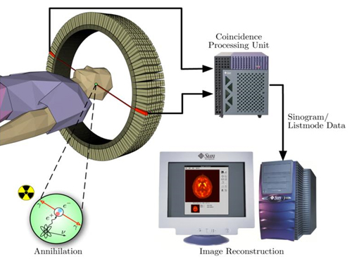
Figure 8. PET detection principle
2013.11.28 View 13884