OT
-
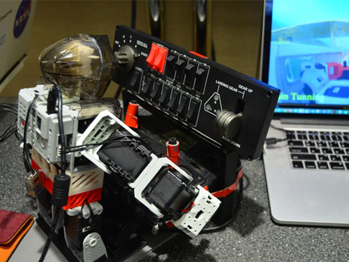 PIBOT, a small humanoid robot flies an aircraft
The 2014 IEEE/RSJ International Conference on Intelligent Robots and Systems (IROS 2014) took place in Chicago, Illinois, on September 14-18, 2014.
Professor David Hyunchul Shim and his students from the Department of Aerospace Engineering, KAIST, presented a research paper entitled “A Robot-machine Interface for Full-functionality Automation Using a Humanoid” at the conference.
The robot called “PIBOT,” a pint-sized, tiny humanoid robot, uses a mixture of flight data and visuals to fly an airplane, capable of identifying and operating all of the buttons and switches in the cockpit of a normal light aircraft designed for humans.
For now, the robot is only flying a simulator, but Professor Shim expects that “PIBOT will help us have a fully automated flight experience, eventually replacing human pilots.”
The IEEE Spectrum magazine published an article on PIBOT posted online September 18, 2014. Please follow the link below for the article:
IEEE Spectrum, September 18, 2014
Tiny Humanoid Robot Learning to Fly Real Airplanes
http://spectrum.ieee.org/automaton/robotics/humanoids/tiny-humanoid-robot-learning-to-fly-real-airplanes
2014.09.23 View 15153
PIBOT, a small humanoid robot flies an aircraft
The 2014 IEEE/RSJ International Conference on Intelligent Robots and Systems (IROS 2014) took place in Chicago, Illinois, on September 14-18, 2014.
Professor David Hyunchul Shim and his students from the Department of Aerospace Engineering, KAIST, presented a research paper entitled “A Robot-machine Interface for Full-functionality Automation Using a Humanoid” at the conference.
The robot called “PIBOT,” a pint-sized, tiny humanoid robot, uses a mixture of flight data and visuals to fly an airplane, capable of identifying and operating all of the buttons and switches in the cockpit of a normal light aircraft designed for humans.
For now, the robot is only flying a simulator, but Professor Shim expects that “PIBOT will help us have a fully automated flight experience, eventually replacing human pilots.”
The IEEE Spectrum magazine published an article on PIBOT posted online September 18, 2014. Please follow the link below for the article:
IEEE Spectrum, September 18, 2014
Tiny Humanoid Robot Learning to Fly Real Airplanes
http://spectrum.ieee.org/automaton/robotics/humanoids/tiny-humanoid-robot-learning-to-fly-real-airplanes
2014.09.23 View 15153 -
 Distinguished Professor Sang Yup Lee Participates in the 2014 Summer Davos Forum
Distinguished Professor Sang Yup Lee from the Department of Chemical and Biomolecular Engineering, KAIST, was invited to lead four sessions at the Annual Meeting 2014, the World Economic Forum, also known as the Summer Davos Forum, which was held in Tianjin, China, from September 10th to 12th.
Two of the four sessions Professor Lee participated in were held on September 10th. At the first session entitled “Biotechnology Ecosystem,” he examined with other panelists the future of bioengineering in depth and discussed major policies and industry trends that will be necessary for the development of future biotechnologies.
Professor Lee later attended the “Strategic Shifts in Healthcare” session as a moderator. Issues related to transforming the health industry such as the next-generation genomics, mobile health and telemedicine, and wearable devices and predictive analytics were addressed.
On September 12, Professor Lee joined the “IdeasLab with KAIST” and gave a presentation on nanotechnology. There was a total of ten IdeasLab sessions held at the Summer Davos Forum, and KAIST was the only Korean university ever invited to host this session. In addition to Professor Lee’s presentation, three more presentations were made by KAIST professors on such topics as “Sustainable Energy and Materials” and “Next-generation Semiconductors.”
Lastly, Professor Lee participated in the “Global Promising Technology” session with the World Economic Forum’s Global Agenda Council members. At this session, he explained the selection of the “World’s Top 10 Most Promising Technologies” and “Bio Sector’s Top 10 Technologies” and led discussions about the “2015 Top 10 Technologies” with the council members.
The Davos Forum has been announcing the “World’s Top 10 Most Promising Technologies” since 2012, and Professor Lee has played a key role in the selection while working as the Chairman of Global Agenda Council. The selection results are presented at the Davos Forum every year and have attracted a lot of attention from around the world.
2014.09.15 View 13789
Distinguished Professor Sang Yup Lee Participates in the 2014 Summer Davos Forum
Distinguished Professor Sang Yup Lee from the Department of Chemical and Biomolecular Engineering, KAIST, was invited to lead four sessions at the Annual Meeting 2014, the World Economic Forum, also known as the Summer Davos Forum, which was held in Tianjin, China, from September 10th to 12th.
Two of the four sessions Professor Lee participated in were held on September 10th. At the first session entitled “Biotechnology Ecosystem,” he examined with other panelists the future of bioengineering in depth and discussed major policies and industry trends that will be necessary for the development of future biotechnologies.
Professor Lee later attended the “Strategic Shifts in Healthcare” session as a moderator. Issues related to transforming the health industry such as the next-generation genomics, mobile health and telemedicine, and wearable devices and predictive analytics were addressed.
On September 12, Professor Lee joined the “IdeasLab with KAIST” and gave a presentation on nanotechnology. There was a total of ten IdeasLab sessions held at the Summer Davos Forum, and KAIST was the only Korean university ever invited to host this session. In addition to Professor Lee’s presentation, three more presentations were made by KAIST professors on such topics as “Sustainable Energy and Materials” and “Next-generation Semiconductors.”
Lastly, Professor Lee participated in the “Global Promising Technology” session with the World Economic Forum’s Global Agenda Council members. At this session, he explained the selection of the “World’s Top 10 Most Promising Technologies” and “Bio Sector’s Top 10 Technologies” and led discussions about the “2015 Top 10 Technologies” with the council members.
The Davos Forum has been announcing the “World’s Top 10 Most Promising Technologies” since 2012, and Professor Lee has played a key role in the selection while working as the Chairman of Global Agenda Council. The selection results are presented at the Davos Forum every year and have attracted a lot of attention from around the world.
2014.09.15 View 13789 -
 News Article on the Development of Synthesis Process for Graphene Quantum Dots
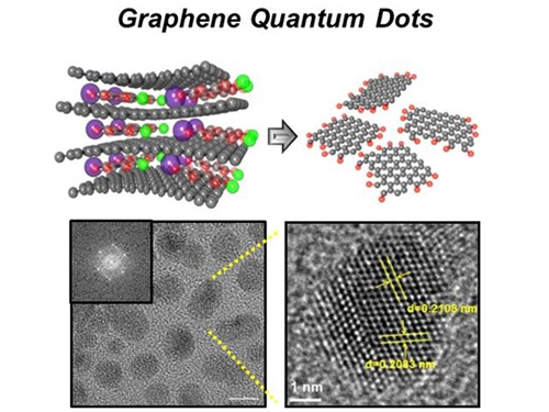
Before It's News, an international online news agency, highlighted the recent research conducted by KAIST professors (Seokwoo Jeon of the Department of Materials Science and Engineering, Yong-Hoon Cho of the Department of Physics, and Seunghyup Yoo of the Department of Electrical Engineering) on the development of synthesis process for graphene quantum dots, nanometer-sized round semiconductor nanoparticles that are very efficient at emitting photons. If commercialized, this synthetic technology will lead the way to the development of paper-thin displays in the future.
For the article, please go to the link below:
Before It’s News, September 3, 2014“Graphene quantum dots prove highly efficient in emitting light”
http://beforeitsnews.com/science-and-technology/2014/09/graphene-quantum-dots-prove-highly-efficient-in-emitting-light-2718190.html
2014.09.07 View 15448
News Article on the Development of Synthesis Process for Graphene Quantum Dots
Before It's News, an international online news agency, highlighted the recent research conducted by KAIST professors (Seokwoo Jeon of the Department of Materials Science and Engineering, Yong-Hoon Cho of the Department of Physics, and Seunghyup Yoo of the Department of Electrical Engineering) on the development of synthesis process for graphene quantum dots, nanometer-sized round semiconductor nanoparticles that are very efficient at emitting photons. If commercialized, this synthetic technology will lead the way to the development of paper-thin displays in the future.
For the article, please go to the link below:
Before It’s News, September 3, 2014“Graphene quantum dots prove highly efficient in emitting light”
http://beforeitsnews.com/science-and-technology/2014/09/graphene-quantum-dots-prove-highly-efficient-in-emitting-light-2718190.html
2014.09.07 View 15448 -
 Extracting Light from Graphite: Core Technology of Graphene Quantum Dots Display Developed
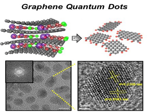
Professor Seokwoo Jeon of the Department of Materials Science and Engineering, Professor Yong-Hoon Cho of the Department of Physics, and Professor Seunghyup Yoo of the Department of Electrical Engineering announced that they were able to develop topnotch graphene quantum dots from graphite.
Using the method of synthesizing graphite intercalation compound from graphite with salt and water, the research team developed graphene quantum dots in an ecofriendly way.
The quantum dots have a diameter of 5 nanometers with their sizes equal and yield high quantum efficiency. Unlike conventional quantum dots, they are not comprised of toxic materials such as lead or cadmium. As the quantum dots can be developed from materials which can be easily found in the nature, researchers look forward to putting these into mass production at low cost.
The research team also discovered a luminescence mechanism of graphene quantum dots and confirmed the possibility of commercial use by developing quantum dot light-emitting diodes with brightness of 1,000 cd/m2, which is greater than that of cellphone displays.
Professor Seokwoo Jeon said, “Although quantum dot LEDs have a lower luminous efficiency than existing ones, their luminescent property can be further improved” and emphasized that “using quantum dot displays will allow us to develop not only paper-thin displays but also flexible ones.”
Sponsored by Graphene Research Center in KAIST Institute for NanoCentury, the research finding was published online in the April 20th issue of Advanced Optical Materials.
Picture 1: Graphene quantum dots and their synthesis
Picture 2: Luminescence mechanism of graphene quantum dots
Picture 3: Structure of graphene quantum dots LED and its emission
2014.09.06 View 19994
Extracting Light from Graphite: Core Technology of Graphene Quantum Dots Display Developed
Professor Seokwoo Jeon of the Department of Materials Science and Engineering, Professor Yong-Hoon Cho of the Department of Physics, and Professor Seunghyup Yoo of the Department of Electrical Engineering announced that they were able to develop topnotch graphene quantum dots from graphite.
Using the method of synthesizing graphite intercalation compound from graphite with salt and water, the research team developed graphene quantum dots in an ecofriendly way.
The quantum dots have a diameter of 5 nanometers with their sizes equal and yield high quantum efficiency. Unlike conventional quantum dots, they are not comprised of toxic materials such as lead or cadmium. As the quantum dots can be developed from materials which can be easily found in the nature, researchers look forward to putting these into mass production at low cost.
The research team also discovered a luminescence mechanism of graphene quantum dots and confirmed the possibility of commercial use by developing quantum dot light-emitting diodes with brightness of 1,000 cd/m2, which is greater than that of cellphone displays.
Professor Seokwoo Jeon said, “Although quantum dot LEDs have a lower luminous efficiency than existing ones, their luminescent property can be further improved” and emphasized that “using quantum dot displays will allow us to develop not only paper-thin displays but also flexible ones.”
Sponsored by Graphene Research Center in KAIST Institute for NanoCentury, the research finding was published online in the April 20th issue of Advanced Optical Materials.
Picture 1: Graphene quantum dots and their synthesis
Picture 2: Luminescence mechanism of graphene quantum dots
Picture 3: Structure of graphene quantum dots LED and its emission
2014.09.06 View 19994 -
 2014 NEREC Conference on Nuclear Nonproliferation: July 31-August 1, 2014, Seoul
The Nonproliferation Education and Research Center (NEREC) at KAIST hosted an international conference on nuclear nonproliferation on July 31-August 1, 2014 in Seoul. The Ministry of Science, ICT and Future Planning, the Korean Nuclear Safety and Security Commission, and the Korea Nuclear Policy Society (KNPS) sponsored the event.
Over one hundred experts and "thought leaders" in nuclear security and nonproliferation attended the conference and discussed issues related to the nonproliferation of nuclear weapons, the role of scientific community in mitigating nuclear threat and promoting the peaceful use of nuclear power, and nuclear disarmament policy.
Keynote speakers were: Steven E. Miller, Director of International Security Program at Belfer Center for Science and International Affairs, Harvard University; Scott D. Sagan, Senior Fellow of the Center for International Security and Cooperation, Freeman Spogli Institute for International Studies, Stanford University; Mark Fitzpatrick, Director of the Nonproliferation and Disarmament Programme, International Institute for Strategic Studies; Sang-Hyun Lee, Director of Security Strategy, Sejong Institute; and Man-Sung Yim, Professor of Nuclear and Quantum Engineering, KAIST.
At the conference, Professor Yim, Director of KAIST NEREC said, “Korea has grown to become a key player in the development of commercial nuclear energy over the past decades. We hope that our conference encourages Korea to be more involved in the efforts of the international community to enhance the global nonproliferation regime.”
2014.08.05 View 16168
2014 NEREC Conference on Nuclear Nonproliferation: July 31-August 1, 2014, Seoul
The Nonproliferation Education and Research Center (NEREC) at KAIST hosted an international conference on nuclear nonproliferation on July 31-August 1, 2014 in Seoul. The Ministry of Science, ICT and Future Planning, the Korean Nuclear Safety and Security Commission, and the Korea Nuclear Policy Society (KNPS) sponsored the event.
Over one hundred experts and "thought leaders" in nuclear security and nonproliferation attended the conference and discussed issues related to the nonproliferation of nuclear weapons, the role of scientific community in mitigating nuclear threat and promoting the peaceful use of nuclear power, and nuclear disarmament policy.
Keynote speakers were: Steven E. Miller, Director of International Security Program at Belfer Center for Science and International Affairs, Harvard University; Scott D. Sagan, Senior Fellow of the Center for International Security and Cooperation, Freeman Spogli Institute for International Studies, Stanford University; Mark Fitzpatrick, Director of the Nonproliferation and Disarmament Programme, International Institute for Strategic Studies; Sang-Hyun Lee, Director of Security Strategy, Sejong Institute; and Man-Sung Yim, Professor of Nuclear and Quantum Engineering, KAIST.
At the conference, Professor Yim, Director of KAIST NEREC said, “Korea has grown to become a key player in the development of commercial nuclear energy over the past decades. We hope that our conference encourages Korea to be more involved in the efforts of the international community to enhance the global nonproliferation regime.”
2014.08.05 View 16168 -
 The First Demonstration of a Self-powered Cardiac Pacemaker
As the number of pacemakers implanted each year reaches into the millions worldwide, improving the lifespan of pacemaker batteries has been of great concern for developers and manufacturers. Currently, pacemaker batteries last seven years on average, requiring frequent replacements, which may pose patients to a potential risk involved in medical procedures.
A research team from the Korea Advanced Institute of Science and Technology (KAIST), headed by Professor Keon Jae Lee of the Department of Materials Science and Engineering at KAIST and Professor Boyoung Joung, M.D. of the Division of Cardiology at Severance Hospital of Yonsei University, has developed a self-powered artificial cardiac pacemaker that is operated semi-permanently by a flexible piezoelectric nanogenerator.
The artificial cardiac pacemaker is widely acknowledged as medical equipment that is integrated into the human body to regulate the heartbeats through electrical stimulation to contract the cardiac muscles of people who suffer from arrhythmia. However, repeated surgeries to replace pacemaker batteries have exposed elderly patients to health risks such as infections or severe bleeding during operations.
The team’s newly designed flexible piezoelectric nanogenerator directly stimulated a living rat’s heart using electrical energy converted from the small body movements of the rat. This technology could facilitate the use of self-powered flexible energy harvesters, not only prolonging the lifetime of cardiac pacemakers but also realizing real-time heart monitoring.
The research team fabricated high-performance flexible nanogenerators utilizing a bulk single-crystal PMN-PT thin film (iBULe Photonics). The harvested energy reached up to 8.2 V and 0.22 mA by bending and pushing motions, which were high enough values to directly stimulate the rat’s heart.
Professor Keon Jae Lee said:
“For clinical purposes, the current achievement will benefit the development of self-powered cardiac pacemakers as well as prevent heart attacks via the real-time diagnosis of heart arrhythmia. In addition, the flexible piezoelectric nanogenerator could also be utilized as an electrical source for various implantable medical devices.”
This research result was described in the April online issue of Advanced Materials (“Self-Powered Cardiac Pacemaker Enabled by Flexible Single Crystalline PMN-PT Piezoelectric Energy Harvester”: http://onlinelibrary.wiley.com/doi/10.1002/adma.201400562/abstract).
Youtube link: http://www.youtube.com/watch?v=ZWYT2cU_Mog&feature=youtu.be
Picture Caption: A self-powered cardiac pacemaker is enabled by a flexible piezoelectric energy harvester.
2014.06.25 View 18163
The First Demonstration of a Self-powered Cardiac Pacemaker
As the number of pacemakers implanted each year reaches into the millions worldwide, improving the lifespan of pacemaker batteries has been of great concern for developers and manufacturers. Currently, pacemaker batteries last seven years on average, requiring frequent replacements, which may pose patients to a potential risk involved in medical procedures.
A research team from the Korea Advanced Institute of Science and Technology (KAIST), headed by Professor Keon Jae Lee of the Department of Materials Science and Engineering at KAIST and Professor Boyoung Joung, M.D. of the Division of Cardiology at Severance Hospital of Yonsei University, has developed a self-powered artificial cardiac pacemaker that is operated semi-permanently by a flexible piezoelectric nanogenerator.
The artificial cardiac pacemaker is widely acknowledged as medical equipment that is integrated into the human body to regulate the heartbeats through electrical stimulation to contract the cardiac muscles of people who suffer from arrhythmia. However, repeated surgeries to replace pacemaker batteries have exposed elderly patients to health risks such as infections or severe bleeding during operations.
The team’s newly designed flexible piezoelectric nanogenerator directly stimulated a living rat’s heart using electrical energy converted from the small body movements of the rat. This technology could facilitate the use of self-powered flexible energy harvesters, not only prolonging the lifetime of cardiac pacemakers but also realizing real-time heart monitoring.
The research team fabricated high-performance flexible nanogenerators utilizing a bulk single-crystal PMN-PT thin film (iBULe Photonics). The harvested energy reached up to 8.2 V and 0.22 mA by bending and pushing motions, which were high enough values to directly stimulate the rat’s heart.
Professor Keon Jae Lee said:
“For clinical purposes, the current achievement will benefit the development of self-powered cardiac pacemakers as well as prevent heart attacks via the real-time diagnosis of heart arrhythmia. In addition, the flexible piezoelectric nanogenerator could also be utilized as an electrical source for various implantable medical devices.”
This research result was described in the April online issue of Advanced Materials (“Self-Powered Cardiac Pacemaker Enabled by Flexible Single Crystalline PMN-PT Piezoelectric Energy Harvester”: http://onlinelibrary.wiley.com/doi/10.1002/adma.201400562/abstract).
Youtube link: http://www.youtube.com/watch?v=ZWYT2cU_Mog&feature=youtu.be
Picture Caption: A self-powered cardiac pacemaker is enabled by a flexible piezoelectric energy harvester.
2014.06.25 View 18163 -
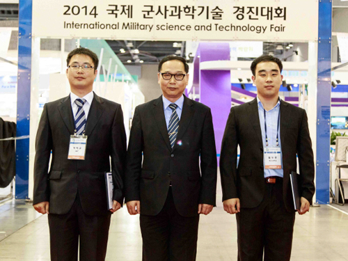 KAIST doctoral student wins prize at 2014 International Military Science and Technology Fair
Min-Kyu Yoo (far left), a doctoral student in the Department of Materials Science Engineering, KAIST, received a silver prize at the 2014 International Military Science and Technology Fair held from May 29 to June 1, 2014 at KINTEX, Ilsan City, Korea.
Yoo presented a paper on aluminum composite materials that were reinforced by carbon nanotubes. Carbon nanotubes reinforced aluminum composite materials have strong mechanical properties, and some nations have used them to manufacture battle tanks.
Aluminum generates hydrogen in an alkaline solution. Utilizing this property and the galvanic corrosion of carbon nanotubes and aluminums, Yoo developed a hydrogen energy system that is fueled with composite materials of carbon nanotube reinforced aluminum. He produced 5 kW electric power and maintained it 22 days using 10 kg of the composite materials for a proton exchange membrane fuel cell and its auxiliary power system.
Yoo’s research will alleviate the difficulty of transporting fuels during wartime and can be applied to the development of an auxiliary power system for next generation aircrafts and battle tanks.
2014.06.24 View 10393
KAIST doctoral student wins prize at 2014 International Military Science and Technology Fair
Min-Kyu Yoo (far left), a doctoral student in the Department of Materials Science Engineering, KAIST, received a silver prize at the 2014 International Military Science and Technology Fair held from May 29 to June 1, 2014 at KINTEX, Ilsan City, Korea.
Yoo presented a paper on aluminum composite materials that were reinforced by carbon nanotubes. Carbon nanotubes reinforced aluminum composite materials have strong mechanical properties, and some nations have used them to manufacture battle tanks.
Aluminum generates hydrogen in an alkaline solution. Utilizing this property and the galvanic corrosion of carbon nanotubes and aluminums, Yoo developed a hydrogen energy system that is fueled with composite materials of carbon nanotube reinforced aluminum. He produced 5 kW electric power and maintained it 22 days using 10 kg of the composite materials for a proton exchange membrane fuel cell and its auxiliary power system.
Yoo’s research will alleviate the difficulty of transporting fuels during wartime and can be applied to the development of an auxiliary power system for next generation aircrafts and battle tanks.
2014.06.24 View 10393 -
 Professor Ki Jun Jeong Selected As the Winner of the 'Young Asian Biotechnologist Prize'
Professor Ki Jun Jeong from the Department of Chemical and Biomolecular Engineering, KAIST, has been selected as the winner of this year’s Young Asian Biotechnologist Prize.
Professor Jeong was invited to the 66th Japan Biotechnology and Bioengineering Society Conference scheduled in September 9th-11th, 2014, in Sapporo, Japan, where his award ceremony will be held.
The award is presented to Professor Jeong in recognition of his outstanding research on microbial-based production of antibodies and efficiency improvement.
The Young Asian Biotechnologist Prize is awarded annually by the Japan Biotechnology and Bioengineering Society to the researchers in Asia under the age of 45, who have achieved excellent research results in the field of bioengineering.
2014.06.14 View 10941
Professor Ki Jun Jeong Selected As the Winner of the 'Young Asian Biotechnologist Prize'
Professor Ki Jun Jeong from the Department of Chemical and Biomolecular Engineering, KAIST, has been selected as the winner of this year’s Young Asian Biotechnologist Prize.
Professor Jeong was invited to the 66th Japan Biotechnology and Bioengineering Society Conference scheduled in September 9th-11th, 2014, in Sapporo, Japan, where his award ceremony will be held.
The award is presented to Professor Jeong in recognition of his outstanding research on microbial-based production of antibodies and efficiency improvement.
The Young Asian Biotechnologist Prize is awarded annually by the Japan Biotechnology and Bioengineering Society to the researchers in Asia under the age of 45, who have achieved excellent research results in the field of bioengineering.
2014.06.14 View 10941 -
 SPIE (The International Society for Optics and Photonics): Scattering Super-lens
The International Society for Optics and Photonics (SPIE), dedicated to advancing an interdisciplinary approach to the science and application of light, published online a short paper authored by a KAIST research team, Dr. Jung-Hoon Park and Professor YongKeun Park of Physics, introducing a new optical technology to observe sub-wavelength light by exploiting multiple light scattering in complex media.
For the article, please go to the link below:
SPIE: Nanotechnology
May 7th, 2014
"Scattering superlens" by Jung-Hoon Park and YongKeun Park
http://spie.org/x108298.xml
2014.05.14 View 8329
SPIE (The International Society for Optics and Photonics): Scattering Super-lens
The International Society for Optics and Photonics (SPIE), dedicated to advancing an interdisciplinary approach to the science and application of light, published online a short paper authored by a KAIST research team, Dr. Jung-Hoon Park and Professor YongKeun Park of Physics, introducing a new optical technology to observe sub-wavelength light by exploiting multiple light scattering in complex media.
For the article, please go to the link below:
SPIE: Nanotechnology
May 7th, 2014
"Scattering superlens" by Jung-Hoon Park and YongKeun Park
http://spie.org/x108298.xml
2014.05.14 View 8329 -
 Clear Display Technology Under Sunlight Developed
The late Professor Seung-Man Yang
The last paper of the late Professor Seung-Man Yang, who was a past master of colloids and fluid mechanics
Practical patterning technology of the next generation optical materials, photonic crystals
The mineral opal does not possess any pigments, but it appears colorful to our eyes. This is because only a particular wavelength is reflected due to the regular nano-structure of its surface. The material that causes selective reflection of the light is called photonic crystals.
The deceased Professor Seung-Man Yang and his research team from KAIST’s Chemical and Biomolecular Engineering Department ha ve developed micro-pattern technology using photolithographic process. This can accelerate the commercialization of photonic crystals, which is hailed as the next generation optics material.
The research results were published in the April 16th edition of Advanced Materials, known as the most prestigious world-renowned journal in the field of materials science.
The newly developed photonic crystal micro-pattern could be used as a core material for the next generation reflective display that is clearly visible even under sunlight. Since it does not require a separate light source, a single charge is enough to last for several days.
Until now, many scientists have endeavored to make photonic crystals artificially, however, most were produced in a lump and therefore lacked efficiency. Also, the low mechanical stability of the formed structure prevented from commercialization.
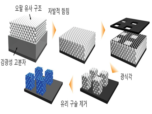
In order to solve these problems, the research team has copied the nano-structure of opals.
Glass beads were arranged in the same nano-structure as the opal on top of the photoresist material undergoing photocuring by ultraviolet light. The glass beads were installed in the photoresist materials, and UV light was selectively exposed on micro regions. The remaining region was developed by photolithographic process to successfully produce photonic crystals in micro-patterns.
The co-author of the research, KAIST Chemical and Biomolecular Engineering Department’s Professor Sin-Hyeon Kim, said, “Combining the semiconductor process technology with photonic crystal pattern technology can secure the practical applications for photonic crystals.”He also predicted “This technology can be used as the key optical material that configures the next generation reflective color display device with very low power consumption.”
The late Professor Seung-Man Yang was a world-renowned expert in the field of colloids and fluid mechanics. Professor Yang published over 193 papers in international journals and continued his research until his passing in last September.
He received Du Pont Science and Technology Award in 2007, KAIST Person of the Year 2008, Gyeong-Am Academy Award in 2009, as well as the President’s Award of the Republic of Korea in March 2014. The researchers devoted the achievement of this year’s research to Professor Yang in his honor.
Research was conducted by KAIST Photonic-fluidic Integrated Devices Research Team, as a part of the Creative Research Program funded by the Ministry of Science, ICT and Future Planning, Republic of Korea.
Figure 1. Opal [left] and the nano glass bead arrangement structure within the opal [right]
Figure 2. Process chart of the photonic crystal micro-pattern formation based on photolithography
Figure 3. Opal structure [left] and inverted structure of the opal [right]
Figure 4. Photonic crystal micro-pattern in solid colors
Figure 5. Photonic crystal micro-pattern that reflects two different crystals (Red, Green) [left] and pixelated pattern of photonic crystal in three primary colors (Red, Green, Blue) [right] that is applicable to reflective displays
2014.05.14 View 14137
Clear Display Technology Under Sunlight Developed
The late Professor Seung-Man Yang
The last paper of the late Professor Seung-Man Yang, who was a past master of colloids and fluid mechanics
Practical patterning technology of the next generation optical materials, photonic crystals
The mineral opal does not possess any pigments, but it appears colorful to our eyes. This is because only a particular wavelength is reflected due to the regular nano-structure of its surface. The material that causes selective reflection of the light is called photonic crystals.
The deceased Professor Seung-Man Yang and his research team from KAIST’s Chemical and Biomolecular Engineering Department ha ve developed micro-pattern technology using photolithographic process. This can accelerate the commercialization of photonic crystals, which is hailed as the next generation optics material.
The research results were published in the April 16th edition of Advanced Materials, known as the most prestigious world-renowned journal in the field of materials science.
The newly developed photonic crystal micro-pattern could be used as a core material for the next generation reflective display that is clearly visible even under sunlight. Since it does not require a separate light source, a single charge is enough to last for several days.
Until now, many scientists have endeavored to make photonic crystals artificially, however, most were produced in a lump and therefore lacked efficiency. Also, the low mechanical stability of the formed structure prevented from commercialization.
In order to solve these problems, the research team has copied the nano-structure of opals.
Glass beads were arranged in the same nano-structure as the opal on top of the photoresist material undergoing photocuring by ultraviolet light. The glass beads were installed in the photoresist materials, and UV light was selectively exposed on micro regions. The remaining region was developed by photolithographic process to successfully produce photonic crystals in micro-patterns.
The co-author of the research, KAIST Chemical and Biomolecular Engineering Department’s Professor Sin-Hyeon Kim, said, “Combining the semiconductor process technology with photonic crystal pattern technology can secure the practical applications for photonic crystals.”He also predicted “This technology can be used as the key optical material that configures the next generation reflective color display device with very low power consumption.”
The late Professor Seung-Man Yang was a world-renowned expert in the field of colloids and fluid mechanics. Professor Yang published over 193 papers in international journals and continued his research until his passing in last September.
He received Du Pont Science and Technology Award in 2007, KAIST Person of the Year 2008, Gyeong-Am Academy Award in 2009, as well as the President’s Award of the Republic of Korea in March 2014. The researchers devoted the achievement of this year’s research to Professor Yang in his honor.
Research was conducted by KAIST Photonic-fluidic Integrated Devices Research Team, as a part of the Creative Research Program funded by the Ministry of Science, ICT and Future Planning, Republic of Korea.
Figure 1. Opal [left] and the nano glass bead arrangement structure within the opal [right]
Figure 2. Process chart of the photonic crystal micro-pattern formation based on photolithography
Figure 3. Opal structure [left] and inverted structure of the opal [right]
Figure 4. Photonic crystal micro-pattern in solid colors
Figure 5. Photonic crystal micro-pattern that reflects two different crystals (Red, Green) [left] and pixelated pattern of photonic crystal in three primary colors (Red, Green, Blue) [right] that is applicable to reflective displays
2014.05.14 View 14137 -
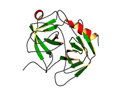 Binding Regulatory Mechanism of Protein Biomolecules Revealed
Professor Hak-Sung Kim
A research team led by Professor Hak-Sung Kim of Biological Sciences, KAIST, and Dr. Mun-Hyeong Seo, KAIST, has revealed a regulatory mechanism that controls the binding affinity of protein’s biomolecules, which is crucial for the protein to recognize molecules and carry out functions within the body.
The research results were published in the April 24th online edition of Nature Communications.
The protein, represented by enzyme, antibody, or hormones, specifically recognizes a variety of biomolecules in all organisms and implements signaling or immune response to precisely adjust and maintain important biological processes. The protein binding affinity of biomolecules plays a crucial role in determining the duration of the bond between two molecules, and hence to determine and control the in-vivo function of proteins.
The researchers have noted that, during the process of proteins’ recognizing biomolecules, the protein binding affinity of biomolecules is closely linked not only to the size of non-covalent interaction between two molecules, but also to the unique kinetic properties of proteins.
To identify the basic mechanism that determines the protein binding affinity of biomolecules, Professor Kim and his research team have made mutation in the allosteric site of protein to create a variety of mutant proteins with the same chemical binding surface, but with the binding affinity vastly differing from 10 to 100 times. The allosteric site of the protein refers to a region which does not directly bind with biomolecules, but crucially influences the biomolecule recognition site.
Using real-time analysis at the single-molecule level of unique kinetic properties of the produced mutant proteins, the researchers were able to identify that the protein binding affinity of biomolecules is directly associated with the protein’s specific kinetic characteristics, its structure opening rate.
Also, by proving that unique characteristics of the protein can be changed at the allosteric site, instead of protein’s direct binding site with biomolecules, the researchers have demonstrated a new methodology of regulating the in-vivo function of proteins.
The researchers expect that these results will contribute greatly to a deeper understanding of protein’s nature that governs various life phenomena and help evaluate the proof of interpreting protein binding affinity of biomolecules from the perspective of protein kinetics.
Professor Kim said, “Until now, the protein binding affinity of biomolecules was determined by a direct interaction between two molecules. Our research has identified an important fact that the structure opening rate of proteins also plays a crucial role in determining their binding affinity.”
[Picture]
A correlation graph of opening rate (kopening) and binding affinity (kd) between protein’s stable, open state and its unstable, partially closed state.
2014.05.02 View 10985
Binding Regulatory Mechanism of Protein Biomolecules Revealed
Professor Hak-Sung Kim
A research team led by Professor Hak-Sung Kim of Biological Sciences, KAIST, and Dr. Mun-Hyeong Seo, KAIST, has revealed a regulatory mechanism that controls the binding affinity of protein’s biomolecules, which is crucial for the protein to recognize molecules and carry out functions within the body.
The research results were published in the April 24th online edition of Nature Communications.
The protein, represented by enzyme, antibody, or hormones, specifically recognizes a variety of biomolecules in all organisms and implements signaling or immune response to precisely adjust and maintain important biological processes. The protein binding affinity of biomolecules plays a crucial role in determining the duration of the bond between two molecules, and hence to determine and control the in-vivo function of proteins.
The researchers have noted that, during the process of proteins’ recognizing biomolecules, the protein binding affinity of biomolecules is closely linked not only to the size of non-covalent interaction between two molecules, but also to the unique kinetic properties of proteins.
To identify the basic mechanism that determines the protein binding affinity of biomolecules, Professor Kim and his research team have made mutation in the allosteric site of protein to create a variety of mutant proteins with the same chemical binding surface, but with the binding affinity vastly differing from 10 to 100 times. The allosteric site of the protein refers to a region which does not directly bind with biomolecules, but crucially influences the biomolecule recognition site.
Using real-time analysis at the single-molecule level of unique kinetic properties of the produced mutant proteins, the researchers were able to identify that the protein binding affinity of biomolecules is directly associated with the protein’s specific kinetic characteristics, its structure opening rate.
Also, by proving that unique characteristics of the protein can be changed at the allosteric site, instead of protein’s direct binding site with biomolecules, the researchers have demonstrated a new methodology of regulating the in-vivo function of proteins.
The researchers expect that these results will contribute greatly to a deeper understanding of protein’s nature that governs various life phenomena and help evaluate the proof of interpreting protein binding affinity of biomolecules from the perspective of protein kinetics.
Professor Kim said, “Until now, the protein binding affinity of biomolecules was determined by a direct interaction between two molecules. Our research has identified an important fact that the structure opening rate of proteins also plays a crucial role in determining their binding affinity.”
[Picture]
A correlation graph of opening rate (kopening) and binding affinity (kd) between protein’s stable, open state and its unstable, partially closed state.
2014.05.02 View 10985 -
 The First Winner of Sang Soo Lee Award in Optics and Photonics
The Optical Society of Korea and the Optical Society of America selected Mario Garavaglia, a researcher at the La Plata Optical Research Center in Argentina, as the first winner of the Sang Soo Lee Award.
Dr. Garavaglia has been selected to receive the award in recognition for his research and education in the field of optics and photonics in Argentina.
The Sang Soo Lee Award, co-established by the Optical Society of Korea and the Optical Society of America in 2012, is awarded to an individual who has made a significant impact in the field. Special considerations are made for individuals who have introduced a new field of research, helped establish a new industry, or made a great contribution to education in the field.
The award is sponsored by the late Doctor Sang Soo Lee's family, the Optical Society of Korea, and the Optical Society of America.
The late Doctor Sang Soo Lee (1925~2010) has been widely known as the 'father of optics' in Korea. He was an active educator, researcher, and writer. Dr. Lee served as the first director of the Korea Advanced Institute of Science (KAIS), the predecessor to KAIST, which was Korea's first research oriented university.
Dr. Lee also served as the 6th president of KAIST between 1989 to 1991 and was a KAIST professor of physics for 21 years. He oversaw the completion of 50 Ph.D. and 100 Master's students as well as published 230 research papers.
Philip Bucksbaum, the president of the Optical Society of America, commented,
"Garavaglia has been an example to the spirit of the Sang Soo Lee Award. The award is the recognition for his tireless efforts and commitment to the development of optics and photonics in Argentina through his teaching, research, and publications."
Jeong-Won Woo, the president of the Optical Society of Korea, said,
"The Sang Soo Lee Award is given to researchers who have consistently contributed to the development of the field. Garavaglia is a well respected researcher in Argentina, and we are truly happy with his selection."
Dr. Garavaglia established a spectroscopy, optic, and laser laboratory in Universidad Nacional de La Plata in 1966. He founded the Center for Optical Research in 1977 and served as the chief of the laboratory until 1991.
Dr. Garavaglia published over 250 research papers in the fields of classical optics, modern optics, photoemission spectroscopy, and laser spectroscopy. He has also received the Galileo Galilei Award from the International Commission for Optics in 1999.
2014.03.31 View 12019
The First Winner of Sang Soo Lee Award in Optics and Photonics
The Optical Society of Korea and the Optical Society of America selected Mario Garavaglia, a researcher at the La Plata Optical Research Center in Argentina, as the first winner of the Sang Soo Lee Award.
Dr. Garavaglia has been selected to receive the award in recognition for his research and education in the field of optics and photonics in Argentina.
The Sang Soo Lee Award, co-established by the Optical Society of Korea and the Optical Society of America in 2012, is awarded to an individual who has made a significant impact in the field. Special considerations are made for individuals who have introduced a new field of research, helped establish a new industry, or made a great contribution to education in the field.
The award is sponsored by the late Doctor Sang Soo Lee's family, the Optical Society of Korea, and the Optical Society of America.
The late Doctor Sang Soo Lee (1925~2010) has been widely known as the 'father of optics' in Korea. He was an active educator, researcher, and writer. Dr. Lee served as the first director of the Korea Advanced Institute of Science (KAIS), the predecessor to KAIST, which was Korea's first research oriented university.
Dr. Lee also served as the 6th president of KAIST between 1989 to 1991 and was a KAIST professor of physics for 21 years. He oversaw the completion of 50 Ph.D. and 100 Master's students as well as published 230 research papers.
Philip Bucksbaum, the president of the Optical Society of America, commented,
"Garavaglia has been an example to the spirit of the Sang Soo Lee Award. The award is the recognition for his tireless efforts and commitment to the development of optics and photonics in Argentina through his teaching, research, and publications."
Jeong-Won Woo, the president of the Optical Society of Korea, said,
"The Sang Soo Lee Award is given to researchers who have consistently contributed to the development of the field. Garavaglia is a well respected researcher in Argentina, and we are truly happy with his selection."
Dr. Garavaglia established a spectroscopy, optic, and laser laboratory in Universidad Nacional de La Plata in 1966. He founded the Center for Optical Research in 1977 and served as the chief of the laboratory until 1991.
Dr. Garavaglia published over 250 research papers in the fields of classical optics, modern optics, photoemission spectroscopy, and laser spectroscopy. He has also received the Galileo Galilei Award from the International Commission for Optics in 1999.
2014.03.31 View 12019