%57%6f%6e%20%44%6f%20%48%65%6f
-
 Unraveling the Secret of Cell Movement
<(From left) Professor Won Do Heo (KAIST), Postdoctoral Researcher Heeyoung Lee (KAIST, First Author), Professor Kwang-Hyun Cho (KAIST), Professor Kapsang Lee (Johns Hopkins University, USA), Dr. Sangkyu Lee (IBS), Dr. Dongsan Kim (LIBD), Dr. Yeaji Seo (Hulux) (Co-First Authors)>
Cell movement is an essential biological process, whether it's cancer cells metastasizing to other parts of the body or immune cells migrating to heal a wound. However, the principle by which cells autonomously determine their direction of movement without external stimuli has remained unknown until now.
Through this research, KAIST and an international joint research team have elucidated the principle by which cells decide their direction and move on their own, offering a crucial clue for identifying the causes of cancer metastasis and immune diseases and establishing new treatment strategies.
KAIST announced on the 10th of November that the research team led by Endowed Chair Professor Won Do Heo of the Department of Biological Sciences, in collaboration with the research team of Endowed Chair Professor Kwang-Hyun Cho of the Department of Bio and Brain Engineering, and Professor Kapsang Lee's research team at Johns Hopkins University in the US, has for the first time in the world identified the 'autonomous driving mechanism' by which cells determine their direction of movement without external signals.
The research team developed a new imaging technique called 'INSPECT (INtracellular Separation of Protein Engineered Condensation Technique)' that allows direct visualization of how proteins interact within living cells. Using this technology, they revealed the principle of the cell's internal program for autonomously deciding its direction of movement.
The team newly analyzed the operation of the key proteins that regulate cell movement, the Rho family proteins (Rac1, Cdc42, RhoA). The results showed that these proteins do not merely divide the front and back of the cell, as previously theorized, but that the cell's decision to move straight or change direction depends on which protein it binds with.
The INSPECT technology artificially implements the phenomenon of 'phase separation,' where proteins, upon binding, naturally form segregated regions that do not mix well. This technique allows for the direct visualization of how proteins actually bind within the cell using a fluorescent signal.
<Figure 1. INSPECT: A technique for visualizing Intracellular Protein-Protein Interactions">
The research team used the proteins ferritin and the fluorescent protein DsRed to make the clusters, or 'condensates,' visible to the eye when proteins bind together like small droplets.
Using this technology, the team analyzed a total of 285 pairs of interactions by combining 15 types of Rho proteins with 19 types of binding proteins, confirming actual binding in 139 pairs. Specifically, they identified that the Cdc42–FMNL protein combination is the core circuit responsible for the cell's 'straight movement,' while the Rac1–ROCK protein combination is responsible for the cell's 'change of direction.'
The research team slightly modified a part of the Rac1 protein (the 37th amino acid), which is crucial for cell direction control, to prevent it from binding well with the 'steering wheel' protein, ROCK. As a result, the cells could not change direction and continued to move in a straight line.
In contrast, in normal cells, Rac1 and ROCK bind well, forming a structure called 'arc stress fiber' at the front of the cell. This fiber enables the cell to make near-perpendicular turns when changing direction.
Furthermore, in an experiment where the environment the cells were attached to was changed, normal cells adjusted their moving speed according to the surrounding environment, but the Rac1F37W cells (cells with a broken 'steering wheel') maintained the same speed regardless of environmental changes. This demonstrates that the Rac–ROCK protein axis subtly controls the cell's ability to recognize and adapt to its surrounding environment.
<Figure 2. Analysis of the Signaling Network through Screening of Protein Interactions that Bind to a Cell Migration-Controlling Protein>
Professor Won Do Heo stated, "This research reveals that cell movement is not a random motion but is precisely controlled by an intrinsic program created by the ensemble of Rho signaling proteins and cell migration-related proteins." He added, "The newly developed INSPECT technology is a powerful tool for visualizing intracellular protein interactions and will be broadly utilized to uncover the molecular mechanisms of various life phenomena and diseases, such as cancer metastasis and neuronal cell migration."
This research, in which Dr. Heeyoung Lee of KAIST, Dr. Sangkyu Lee (currently at IBS), Dr. Yeji Seo (currently at Hulux Co., Ltd.), and Dr. Dongsan Kim (currently at LIBD) participated as co-first authors, was published in Nature Communications on October 31st.
Journal Name: A Rho GTPase-effector ensemble governs cell migration behavior
DOI: https://doi.org/10.1038/s41467-025-64635-0
The research was supported by the Samsung Future Technology Foundation and the National Research Foundation of Korea.
2025.11.10 View 2010
Unraveling the Secret of Cell Movement
<(From left) Professor Won Do Heo (KAIST), Postdoctoral Researcher Heeyoung Lee (KAIST, First Author), Professor Kwang-Hyun Cho (KAIST), Professor Kapsang Lee (Johns Hopkins University, USA), Dr. Sangkyu Lee (IBS), Dr. Dongsan Kim (LIBD), Dr. Yeaji Seo (Hulux) (Co-First Authors)>
Cell movement is an essential biological process, whether it's cancer cells metastasizing to other parts of the body or immune cells migrating to heal a wound. However, the principle by which cells autonomously determine their direction of movement without external stimuli has remained unknown until now.
Through this research, KAIST and an international joint research team have elucidated the principle by which cells decide their direction and move on their own, offering a crucial clue for identifying the causes of cancer metastasis and immune diseases and establishing new treatment strategies.
KAIST announced on the 10th of November that the research team led by Endowed Chair Professor Won Do Heo of the Department of Biological Sciences, in collaboration with the research team of Endowed Chair Professor Kwang-Hyun Cho of the Department of Bio and Brain Engineering, and Professor Kapsang Lee's research team at Johns Hopkins University in the US, has for the first time in the world identified the 'autonomous driving mechanism' by which cells determine their direction of movement without external signals.
The research team developed a new imaging technique called 'INSPECT (INtracellular Separation of Protein Engineered Condensation Technique)' that allows direct visualization of how proteins interact within living cells. Using this technology, they revealed the principle of the cell's internal program for autonomously deciding its direction of movement.
The team newly analyzed the operation of the key proteins that regulate cell movement, the Rho family proteins (Rac1, Cdc42, RhoA). The results showed that these proteins do not merely divide the front and back of the cell, as previously theorized, but that the cell's decision to move straight or change direction depends on which protein it binds with.
The INSPECT technology artificially implements the phenomenon of 'phase separation,' where proteins, upon binding, naturally form segregated regions that do not mix well. This technique allows for the direct visualization of how proteins actually bind within the cell using a fluorescent signal.
<Figure 1. INSPECT: A technique for visualizing Intracellular Protein-Protein Interactions">
The research team used the proteins ferritin and the fluorescent protein DsRed to make the clusters, or 'condensates,' visible to the eye when proteins bind together like small droplets.
Using this technology, the team analyzed a total of 285 pairs of interactions by combining 15 types of Rho proteins with 19 types of binding proteins, confirming actual binding in 139 pairs. Specifically, they identified that the Cdc42–FMNL protein combination is the core circuit responsible for the cell's 'straight movement,' while the Rac1–ROCK protein combination is responsible for the cell's 'change of direction.'
The research team slightly modified a part of the Rac1 protein (the 37th amino acid), which is crucial for cell direction control, to prevent it from binding well with the 'steering wheel' protein, ROCK. As a result, the cells could not change direction and continued to move in a straight line.
In contrast, in normal cells, Rac1 and ROCK bind well, forming a structure called 'arc stress fiber' at the front of the cell. This fiber enables the cell to make near-perpendicular turns when changing direction.
Furthermore, in an experiment where the environment the cells were attached to was changed, normal cells adjusted their moving speed according to the surrounding environment, but the Rac1F37W cells (cells with a broken 'steering wheel') maintained the same speed regardless of environmental changes. This demonstrates that the Rac–ROCK protein axis subtly controls the cell's ability to recognize and adapt to its surrounding environment.
<Figure 2. Analysis of the Signaling Network through Screening of Protein Interactions that Bind to a Cell Migration-Controlling Protein>
Professor Won Do Heo stated, "This research reveals that cell movement is not a random motion but is precisely controlled by an intrinsic program created by the ensemble of Rho signaling proteins and cell migration-related proteins." He added, "The newly developed INSPECT technology is a powerful tool for visualizing intracellular protein interactions and will be broadly utilized to uncover the molecular mechanisms of various life phenomena and diseases, such as cancer metastasis and neuronal cell migration."
This research, in which Dr. Heeyoung Lee of KAIST, Dr. Sangkyu Lee (currently at IBS), Dr. Yeji Seo (currently at Hulux Co., Ltd.), and Dr. Dongsan Kim (currently at LIBD) participated as co-first authors, was published in Nature Communications on October 31st.
Journal Name: A Rho GTPase-effector ensemble governs cell migration behavior
DOI: https://doi.org/10.1038/s41467-025-64635-0
The research was supported by the Samsung Future Technology Foundation and the National Research Foundation of Korea.
2025.11.10 View 2010 -
 A Breakthrough in Parkinson's Research: Precision Diagnosis and Treatment with AI and Optogenetics
<Research team photo (from top left) Dr. Bobae Hyeon, Professor Daesoo Kim, Director Chang-joon Lee, (right) Professor Won Do Heo>
Globally recognized figures like Muhammad Ali and Michael J. Fox have long suffered from Parkinson's disease. The disease presents a complex set of motor symptoms, including tremors, rigidity, bradykinesia, and postural instability. However, traditional diagnostic methods have struggled to sensitively detect changes in the early stages, and drugs targeting brain signal regulation have had limited clinical effectiveness.
Recently, Korean researchers successfully demonstrated the potential of a technology that integrates AI and optogenetics as a tool for precise diagnosis and therapeutic evaluation of Parkinson's disease in mice. They have also proposed a strategy for developing next-generation personalized treatments.
KAIST (President Kwang Hyung Lee) announced on the 22nd of September that a collaborative research team—comprising Professor Won Do Heo's team from the Department of Biological Sciences, Professor Daesoo Kim's team from the Department of Brain and Cognitive Sciences, and Director Chang-Jun Lee's team from the Institute for Basic Science (IBS) Center for Cognition and Sociality—achieved a preclinical research breakthrough by combining AI analysis with optogenetics. Their work simultaneously demonstrated the possibility of early and precise diagnosis and treatment in an animal model of Parkinson's disease.
The research team created a Parkinson's disease mouse model with two stages of severity. These were male mice with alpha-synuclein protein abnormalities, a standard model used to simulate human Parkinson's disease for diagnostic and therapeutic research.
In collaboration with Professor Kim's team at KAIST, they introduced AI-based 3D pose estimation for behavioral analysis. The team analyzed over 340 behavioral features—such as gait, limb movements, and tremors—from the Parkinson's mice and condensed them into a single metric: the AI-predicted Parkinson's disease score (APS).
The analysis showed that the APS exhibited a significant difference from the control group as early as two weeks after the disease was induced. It also proved more sensitive in assessing the disease's severity than traditional motor function tests. The study identified key diagnostic features, including changes in stride, asymmetrical limb movements, and chest tremors. The top 20 behavioral features included hand/foot asymmetry, changes in stride and posture, and an increase in high-frequency chest movement.
To confirm that these behavioral indicators were not just general motor decline but specific to Parkinson's, the team applied the same analysis to a mouse model of Amyotrophic Lateral Sclerosis (ALS), also known as Lou Gehrig's disease, in collaboration with Director Lee's team at IBS. Since both Parkinson's and ALS cause motor function problems, if the APS simply reflected poor motor skills, a high score should have appeared in both diseases.
However, the analysis of the ALS animal model showed that despite a decline in motor function, the mice did not exhibit the high APS seen in the Parkinson's model. Instead, their scores remained low, and their behavioral changes were distinctly different. This demonstrates that APS is directly related to specific, characteristic changes that only appear in Parkinson's disease.
For treatment, the research team used optoRET, an optogenetics technology that precisely controls neurotrophic signals with light. This technique proved effective in the animal model, leading to smoother gait and limb movements and a reduction in tremors.
Specifically, a regimen of shining light on alternate days was found to be the most effective, and it also showed a tendency to protect dopamine-producing neurons in the brain.
Professor Won Do Heo of KAIST stated, "This is the first time in the world that a preclinical framework has been implemented that connects early diagnosis, treatment evaluation, and mechanism verification of Parkinson's disease by combining AI-based behavioral analysis with optogenetics." He added, "This lays a crucial foundation for future personalized medicine and customized treatments for patients."
The study, with Dr. Bobae Hyeon, a postdoctoral researcher at the KAIST Institute for Biological Science, as the first author, was published online in the international journal Nature Communications on August 21st. Dr. Hyeon is conducting follow-up research to advance Parkinson's cell therapy at McLean Hospital, Harvard Medical School, supported by the "Global Physician-Scientist Training Program" of the Korea Health Industry Development Institute.
This research was supported by the KAIST Global Singularity Project, the Ministry of Science and ICT/National Research Foundation of Korea, the IBS Center for Cognition and Sociality, and the Ministry of Health and Welfare/Korea Health Industry Development Institute.
Paper Title: Integrating artificial intelligence and optogenetics for Parkinson's disease diagnosis and therapeutics in male mice
DOI: https://doi.org/10.1038/s41467-025-63025-w
2025.09.22 View 2546
A Breakthrough in Parkinson's Research: Precision Diagnosis and Treatment with AI and Optogenetics
<Research team photo (from top left) Dr. Bobae Hyeon, Professor Daesoo Kim, Director Chang-joon Lee, (right) Professor Won Do Heo>
Globally recognized figures like Muhammad Ali and Michael J. Fox have long suffered from Parkinson's disease. The disease presents a complex set of motor symptoms, including tremors, rigidity, bradykinesia, and postural instability. However, traditional diagnostic methods have struggled to sensitively detect changes in the early stages, and drugs targeting brain signal regulation have had limited clinical effectiveness.
Recently, Korean researchers successfully demonstrated the potential of a technology that integrates AI and optogenetics as a tool for precise diagnosis and therapeutic evaluation of Parkinson's disease in mice. They have also proposed a strategy for developing next-generation personalized treatments.
KAIST (President Kwang Hyung Lee) announced on the 22nd of September that a collaborative research team—comprising Professor Won Do Heo's team from the Department of Biological Sciences, Professor Daesoo Kim's team from the Department of Brain and Cognitive Sciences, and Director Chang-Jun Lee's team from the Institute for Basic Science (IBS) Center for Cognition and Sociality—achieved a preclinical research breakthrough by combining AI analysis with optogenetics. Their work simultaneously demonstrated the possibility of early and precise diagnosis and treatment in an animal model of Parkinson's disease.
The research team created a Parkinson's disease mouse model with two stages of severity. These were male mice with alpha-synuclein protein abnormalities, a standard model used to simulate human Parkinson's disease for diagnostic and therapeutic research.
In collaboration with Professor Kim's team at KAIST, they introduced AI-based 3D pose estimation for behavioral analysis. The team analyzed over 340 behavioral features—such as gait, limb movements, and tremors—from the Parkinson's mice and condensed them into a single metric: the AI-predicted Parkinson's disease score (APS).
The analysis showed that the APS exhibited a significant difference from the control group as early as two weeks after the disease was induced. It also proved more sensitive in assessing the disease's severity than traditional motor function tests. The study identified key diagnostic features, including changes in stride, asymmetrical limb movements, and chest tremors. The top 20 behavioral features included hand/foot asymmetry, changes in stride and posture, and an increase in high-frequency chest movement.
To confirm that these behavioral indicators were not just general motor decline but specific to Parkinson's, the team applied the same analysis to a mouse model of Amyotrophic Lateral Sclerosis (ALS), also known as Lou Gehrig's disease, in collaboration with Director Lee's team at IBS. Since both Parkinson's and ALS cause motor function problems, if the APS simply reflected poor motor skills, a high score should have appeared in both diseases.
However, the analysis of the ALS animal model showed that despite a decline in motor function, the mice did not exhibit the high APS seen in the Parkinson's model. Instead, their scores remained low, and their behavioral changes were distinctly different. This demonstrates that APS is directly related to specific, characteristic changes that only appear in Parkinson's disease.
For treatment, the research team used optoRET, an optogenetics technology that precisely controls neurotrophic signals with light. This technique proved effective in the animal model, leading to smoother gait and limb movements and a reduction in tremors.
Specifically, a regimen of shining light on alternate days was found to be the most effective, and it also showed a tendency to protect dopamine-producing neurons in the brain.
Professor Won Do Heo of KAIST stated, "This is the first time in the world that a preclinical framework has been implemented that connects early diagnosis, treatment evaluation, and mechanism verification of Parkinson's disease by combining AI-based behavioral analysis with optogenetics." He added, "This lays a crucial foundation for future personalized medicine and customized treatments for patients."
The study, with Dr. Bobae Hyeon, a postdoctoral researcher at the KAIST Institute for Biological Science, as the first author, was published online in the international journal Nature Communications on August 21st. Dr. Hyeon is conducting follow-up research to advance Parkinson's cell therapy at McLean Hospital, Harvard Medical School, supported by the "Global Physician-Scientist Training Program" of the Korea Health Industry Development Institute.
This research was supported by the KAIST Global Singularity Project, the Ministry of Science and ICT/National Research Foundation of Korea, the IBS Center for Cognition and Sociality, and the Ministry of Health and Welfare/Korea Health Industry Development Institute.
Paper Title: Integrating artificial intelligence and optogenetics for Parkinson's disease diagnosis and therapeutics in male mice
DOI: https://doi.org/10.1038/s41467-025-63025-w
2025.09.22 View 2546 -
 “Why are we depressed?” KAIST is identifying the cause of depression and uncovering clues for treatment
Major depressive disorder (MDD) is one of the most common psychiatric illnesses worldwide, but its molecular causes* have still not been clearly identified. A domestic research team has discovered that depression may not simply be caused by neuronal damage, but can also arise from the dysregulation of specific neural signaling pathways. In particular, they identified the molecular reason why elderly patients with depression do not respond to conventional antidepressants. This study suggests the possibility of therapeutic approaches using optogenetic technology to regulate neural signaling, and it provides clues for the development of new treatment strategies targeting the protein ‘Numb’ protein for elderly patients with depression.
*Molecular causes: explanations for the origin of a disease at the level of molecules, proteins, or genes in the brain.
KAIST (President Kwang Hyung Lee) announced on the 19th of August that a research team led by Distinguished Professor Won Do Heo of the Department of Biological Sciences at KAIST, in collaboration with forensic pathologist Minju Lee of the National Forensic Service (Director Bong Woo Lee) and Professor Seokhwi Kim of the Department of Pathology at Ajou University Medical Center (Director Sangwook Han), identified a new molecular mechanism for depression through RNA sequencing and the immunohistochemical analysis of brain tissue from patients who had committed suicide. Furthermore, they demonstrated in animal models that antidepressant effects can be restored by regulating the signaling pathway that induces neural recovery using optogenetic technology.
The research team focused on the hippocampus, the brain region responsible for memory and emotion, and in particular on the dentate gyrus (DG). The DG is the entry point of information into the hippocampus, playing a role in new memory formation, neurogenesis, and emotional regulation, and is closely linked with depression.
Using two representative mouse models for depression (the corticosterone stress model and the chronic unpredictable stress model), the team found that stress induced a striking increase in the signaling receptor FGFR1 (Fibroblast Growth Factor Receptor 1) in the DG. FGFR1 receives growth factor (FGF) signals and transmits growth and differentiation commands within cells.
Subsequently, using conditional knockout (cKO) mice in which the FGFR1 gene was deleted, the researchers revealed that the absence of FGFR1 made mice more vulnerable to stress and led them to exhibit depressive symptoms more quickly. This indicates that FGFR1 plays a critical role in proper neural regulation and stress resistance.
The team then developed an ‘optoFGFR1 system’ using optogenetics, enabling FGFR1 —essential for stress resistance—to be activated by light. They observed that activating FGFR1 in depression mouse models lacking FGFR1 restored antidepressant effects. In other words, they experimentally demonstrated that the activation of FGFR1 signaling alone could improve depressive behavior.
Surprisingly, however, in aged depression mouse models, the activation of FGFR1 signaling through the optoFGFR1 system did not yield antidepressant effects. Investigating further, the researchers found that in the aged brains, a protein called ‘Numb’ was excessively expressed and interfered with FGFR1 signaling.
Indeed, analysis of postmortem human brain tissue also showed the specific overexpression of Numb protein only in elderly patients with depression. When the researchers suppressed Numb using a gene regulatory tool (shRNA) while simultaneously activating FGFR1 signaling in mouse models, neurogenesis and behavior—previously unrecoverable—returned to normal even in aged depression models. This shows that the Numb protein acts as a “blocker” of FGFR1 signaling and is a key factor preventing the hippocampus from executing antidepressant mechanisms.
Distinguished Professor Won Do Heo of KAIST said, “This study is meaningful in that it revealed that depression may not only result from simple neuronal damage, but can also arise from the dysregulation of specific neural signaling pathways. In particular, we identified the molecular reason why antidepressants are less effective in elderly patients, and we expect this to provide a clue for the development of new therapeutic strategies targeting the Numb protein.”
He added, “Moreover, this interdisciplinary study, which combined KAIST’s expertise in neuroscience with the National Forensic Service’s forensic brain analysis technologies, is expected to serve as a bridge between basic research on psychiatric disorders and clinical applications.”
This study, led by first author Jongpil Shin, a PhD student in the Department of Biological Sciences at KAIST, was published on August 15, 2025, in the international journal Experimental & Molecular Medicine.
Paper title: “Dysregulation of FGFR1 signaling in the hippocampus facilitates depressive disorder”
DOI: https://doi.org/10.1038/s12276-025-01519-9
This research was supported by the Ministry of Science and ICT’s National Research Foundation of Korea through the ASTRA program and the Bio-Medical Technology Development project.
2025.08.19 View 3556
“Why are we depressed?” KAIST is identifying the cause of depression and uncovering clues for treatment
Major depressive disorder (MDD) is one of the most common psychiatric illnesses worldwide, but its molecular causes* have still not been clearly identified. A domestic research team has discovered that depression may not simply be caused by neuronal damage, but can also arise from the dysregulation of specific neural signaling pathways. In particular, they identified the molecular reason why elderly patients with depression do not respond to conventional antidepressants. This study suggests the possibility of therapeutic approaches using optogenetic technology to regulate neural signaling, and it provides clues for the development of new treatment strategies targeting the protein ‘Numb’ protein for elderly patients with depression.
*Molecular causes: explanations for the origin of a disease at the level of molecules, proteins, or genes in the brain.
KAIST (President Kwang Hyung Lee) announced on the 19th of August that a research team led by Distinguished Professor Won Do Heo of the Department of Biological Sciences at KAIST, in collaboration with forensic pathologist Minju Lee of the National Forensic Service (Director Bong Woo Lee) and Professor Seokhwi Kim of the Department of Pathology at Ajou University Medical Center (Director Sangwook Han), identified a new molecular mechanism for depression through RNA sequencing and the immunohistochemical analysis of brain tissue from patients who had committed suicide. Furthermore, they demonstrated in animal models that antidepressant effects can be restored by regulating the signaling pathway that induces neural recovery using optogenetic technology.
The research team focused on the hippocampus, the brain region responsible for memory and emotion, and in particular on the dentate gyrus (DG). The DG is the entry point of information into the hippocampus, playing a role in new memory formation, neurogenesis, and emotional regulation, and is closely linked with depression.
Using two representative mouse models for depression (the corticosterone stress model and the chronic unpredictable stress model), the team found that stress induced a striking increase in the signaling receptor FGFR1 (Fibroblast Growth Factor Receptor 1) in the DG. FGFR1 receives growth factor (FGF) signals and transmits growth and differentiation commands within cells.
Subsequently, using conditional knockout (cKO) mice in which the FGFR1 gene was deleted, the researchers revealed that the absence of FGFR1 made mice more vulnerable to stress and led them to exhibit depressive symptoms more quickly. This indicates that FGFR1 plays a critical role in proper neural regulation and stress resistance.
The team then developed an ‘optoFGFR1 system’ using optogenetics, enabling FGFR1 —essential for stress resistance—to be activated by light. They observed that activating FGFR1 in depression mouse models lacking FGFR1 restored antidepressant effects. In other words, they experimentally demonstrated that the activation of FGFR1 signaling alone could improve depressive behavior.
Surprisingly, however, in aged depression mouse models, the activation of FGFR1 signaling through the optoFGFR1 system did not yield antidepressant effects. Investigating further, the researchers found that in the aged brains, a protein called ‘Numb’ was excessively expressed and interfered with FGFR1 signaling.
Indeed, analysis of postmortem human brain tissue also showed the specific overexpression of Numb protein only in elderly patients with depression. When the researchers suppressed Numb using a gene regulatory tool (shRNA) while simultaneously activating FGFR1 signaling in mouse models, neurogenesis and behavior—previously unrecoverable—returned to normal even in aged depression models. This shows that the Numb protein acts as a “blocker” of FGFR1 signaling and is a key factor preventing the hippocampus from executing antidepressant mechanisms.
Distinguished Professor Won Do Heo of KAIST said, “This study is meaningful in that it revealed that depression may not only result from simple neuronal damage, but can also arise from the dysregulation of specific neural signaling pathways. In particular, we identified the molecular reason why antidepressants are less effective in elderly patients, and we expect this to provide a clue for the development of new therapeutic strategies targeting the Numb protein.”
He added, “Moreover, this interdisciplinary study, which combined KAIST’s expertise in neuroscience with the National Forensic Service’s forensic brain analysis technologies, is expected to serve as a bridge between basic research on psychiatric disorders and clinical applications.”
This study, led by first author Jongpil Shin, a PhD student in the Department of Biological Sciences at KAIST, was published on August 15, 2025, in the international journal Experimental & Molecular Medicine.
Paper title: “Dysregulation of FGFR1 signaling in the hippocampus facilitates depressive disorder”
DOI: https://doi.org/10.1038/s12276-025-01519-9
This research was supported by the Ministry of Science and ICT’s National Research Foundation of Korea through the ASTRA program and the Bio-Medical Technology Development project.
2025.08.19 View 3556 -
 KAIST Team Develops Optogenetic Platform for Spatiotemporal Control of Protein and mRNA Storage and Release
<Dr. Chaeyeon Lee, Professor Won Do Heo from Department of Biological Sciences>
A KAIST research team led by Professor Won Do Heo (Department of Biological Sciences) has developed an optogenetic platform, RELISR (REversible LIght-induced Store and Release), that enables precise spatiotemporal control over the storage and release of proteins and mRNAs in living cells and animals.
Traditional optogenetic condensate systems have been limited by their reliance on non-specific multivalent interactions, which can lead to unintended sequestration or release of endogenous molecules. RELISR overcomes these limitations by employing highly specific protein–protein (nanobody–antigen) and protein–RNA (MCP–MS2) interactions, enabling the selective and reversible compartmentalization of target proteins or mRNAs within engineered, membrane-less condensates.
In the dark, RELISR stably sequesters target molecules within condensates, physically isolating them from the cellular environment. Upon blue light stimulation, the condensates rapidly dissolve, releasing the stored proteins or mRNAs, which immediately regain their cellular functions or translational competency. This allows for reversible and rapid modulation of molecular activities in response to optical cues.
< Figure 1. Overview of the Artificial Condensate System (RELISR). The artificial condensate system, RELISR, includes "Protein-RELISR" for storing proteins and "mRNA-RELISR" for storing mRNA. These artificial condensates can be disassembled by blue light irradiation and reassembled in a dark state>
The research team demonstrated that RELISR enables temporal and spatial regulation of protein activity and mRNA translation in various cell types, including cultured neurons and mouse liver tissue. Comparative studies showed that RELISR provides more robust and reversible control of translation than previous systems based on spatial translocation.
While previous optogenetic systems such as LARIAT (Lee et al., Nature Methods, 2014) and mRNA-LARIAT (Kim et al., Nat. Cell Biol., 2019) enabled the selective sequestration of proteins or mRNAs into membrane-less condensates in response to light, they were primarily limited to the trapping phase. The RELISR platform introduced in this study establishes a new paradigm by enabling both the targeted storage of proteins and mRNAs and their rapid, light-triggered release. This approach allows researchers to not only confine molecular function on demand, but also to restore activity with precise temporal control.
< Figure 2. Cell shape change using the artificial condensate system (RELISR). A target protein, Vav2, which contributes to cell shape, was stored within the artificial condensate and then released after light irradiation. This release activated the target protein Vav2, causing a change in cell shape. It was confirmed that the storage, release, and activation of various proteins were effectively achieved>
Professor Heo stated, “RELISR is a versatile optogenetic tool that enables the precise control of protein and mRNA function at defined times and locations in living systems. We anticipate this platform will be broadly applicable for studies of cell signaling, neural circuits, and therapeutic development. Furthermore, the combination of RELISR with genome editing or tissue-targeted delivery could further expand its utility for molecular medicine.”
< Figure 3. Expression of a target mRNA using the artificial condensate system (RELISR) in mice. The genetic material for the artificial condensate system, RELISR, was injected into a living mouse. Using this system, a target mRNA was stored within the mouse's liver. Upon light irradiation, the mRNA was released, which induced the translation of a luminescent protein>
This research was conducted by first author Dr. Chaeyeon Lee, under the supervision of Professor Heo, with contributions from Dr. Daseuli Yu (co-corresponding author) and Professor YongKeun Park (co-corresponding author, Department of Physics), whose group performed quantitative imaging analyses of biophysical changes induced by RELISR in cells.
The findings were published in Nature Communications (July 7, 2025; DOI: 10.1038/s41467-025-61322-y). This work was supported by the Samsung Future Technology Foundation and the National Research Foundation of Korea.
2025.07.23 View 4114
KAIST Team Develops Optogenetic Platform for Spatiotemporal Control of Protein and mRNA Storage and Release
<Dr. Chaeyeon Lee, Professor Won Do Heo from Department of Biological Sciences>
A KAIST research team led by Professor Won Do Heo (Department of Biological Sciences) has developed an optogenetic platform, RELISR (REversible LIght-induced Store and Release), that enables precise spatiotemporal control over the storage and release of proteins and mRNAs in living cells and animals.
Traditional optogenetic condensate systems have been limited by their reliance on non-specific multivalent interactions, which can lead to unintended sequestration or release of endogenous molecules. RELISR overcomes these limitations by employing highly specific protein–protein (nanobody–antigen) and protein–RNA (MCP–MS2) interactions, enabling the selective and reversible compartmentalization of target proteins or mRNAs within engineered, membrane-less condensates.
In the dark, RELISR stably sequesters target molecules within condensates, physically isolating them from the cellular environment. Upon blue light stimulation, the condensates rapidly dissolve, releasing the stored proteins or mRNAs, which immediately regain their cellular functions or translational competency. This allows for reversible and rapid modulation of molecular activities in response to optical cues.
< Figure 1. Overview of the Artificial Condensate System (RELISR). The artificial condensate system, RELISR, includes "Protein-RELISR" for storing proteins and "mRNA-RELISR" for storing mRNA. These artificial condensates can be disassembled by blue light irradiation and reassembled in a dark state>
The research team demonstrated that RELISR enables temporal and spatial regulation of protein activity and mRNA translation in various cell types, including cultured neurons and mouse liver tissue. Comparative studies showed that RELISR provides more robust and reversible control of translation than previous systems based on spatial translocation.
While previous optogenetic systems such as LARIAT (Lee et al., Nature Methods, 2014) and mRNA-LARIAT (Kim et al., Nat. Cell Biol., 2019) enabled the selective sequestration of proteins or mRNAs into membrane-less condensates in response to light, they were primarily limited to the trapping phase. The RELISR platform introduced in this study establishes a new paradigm by enabling both the targeted storage of proteins and mRNAs and their rapid, light-triggered release. This approach allows researchers to not only confine molecular function on demand, but also to restore activity with precise temporal control.
< Figure 2. Cell shape change using the artificial condensate system (RELISR). A target protein, Vav2, which contributes to cell shape, was stored within the artificial condensate and then released after light irradiation. This release activated the target protein Vav2, causing a change in cell shape. It was confirmed that the storage, release, and activation of various proteins were effectively achieved>
Professor Heo stated, “RELISR is a versatile optogenetic tool that enables the precise control of protein and mRNA function at defined times and locations in living systems. We anticipate this platform will be broadly applicable for studies of cell signaling, neural circuits, and therapeutic development. Furthermore, the combination of RELISR with genome editing or tissue-targeted delivery could further expand its utility for molecular medicine.”
< Figure 3. Expression of a target mRNA using the artificial condensate system (RELISR) in mice. The genetic material for the artificial condensate system, RELISR, was injected into a living mouse. Using this system, a target mRNA was stored within the mouse's liver. Upon light irradiation, the mRNA was released, which induced the translation of a luminescent protein>
This research was conducted by first author Dr. Chaeyeon Lee, under the supervision of Professor Heo, with contributions from Dr. Daseuli Yu (co-corresponding author) and Professor YongKeun Park (co-corresponding author, Department of Physics), whose group performed quantitative imaging analyses of biophysical changes induced by RELISR in cells.
The findings were published in Nature Communications (July 7, 2025; DOI: 10.1038/s41467-025-61322-y). This work was supported by the Samsung Future Technology Foundation and the National Research Foundation of Korea.
2025.07.23 View 4114 -
 KAIST develops technology for selective RNA modification in living cells and animals
· A team led by Professor Won Do Heo from the Department of Biological Sciences, KAIST, has developed a pioneering technology that selectively acetylates specific RNA molecules in living cells and tissues.
· The platform uses RNA-targeting CRISPR tools in combination with RNA-modifying enzymes to chemically modify only the intended RNA.
· The method opens new possibilities for gene therapy by enabling precise control of disease-related RNA without affecting the rest of the transcriptome.
< Photo 1. (From left) Professor Won Do Heo and Jihwan Yu, a Ph.D. Candidate of the Department of Biological Sciences >
CRISPR-Cas13, a powerful RNA-targeting technology is gaining increasing attention as a next-generation gene therapy platform due to its precision and reduced side effects. Utilizing this system, researchers at KAIST have now developed the world’s first technology capable of selectively acetylating (chemically modifying) specific RNA molecules among countless transcripts within living cells. This breakthrough enables precise, programmable control of RNA function and is expected to open new avenues in RNA-based therapeutic development.
KAIST (President Kwang Hyung Lee) announced that a research team led by Professor Won Do Heo in the Department of Biological Sciences has recently developed a groundbreaking technology capable of selectively acetylating specific RNA molecules within the human body using the CRISPR-Cas13 system—an RNA-targeting platform gaining increasing attention in the fields of gene regulation and RNA-based therapeutics.
RNA molecules can undergo chemical modifications—the addition of specific chemical groups—which alter their function and behavior without changing the underlying nucleotide sequence. However, some of these modifications, a critical layer of post-transcriptional gene regulation, remain poorly understood. Among them, N4-acetylcytidine (ac4C) has been particularly enigmatic, with ongoing debate about its existence and function in human messenger RNA (mRNA), the RNA that encodes proteins.
To address this gap, the KAIST research team developed a targeted RNA acetylation system, named dCas13-eNAT10. This platform combines a catalytically inactive Cas13 enzyme (dCas13) that guides the system to specific RNA targets, with a hyperactive variant of the NAT10 enzyme (eNAT10), which performs RNA acetylation. This approach enables precise acetylation of only the desired RNA molecules among the vast pool of transcripts within the cell.
< Figure 1. Development of hyperactive variant eNAT10 through NAT10 protein engineering. By engineering the NAT10 protein, which performs RNA acetylation in human cells, based on its domain and structure, eNAT10 was developed, showing approximately a 3-fold increase in RNA acetylation activity compared to the wild-type enzyme. >
Using this system, the researchers demonstrated that guide RNAs could direct the dCas13-eNAT10 complex to acetylate specific RNA targets, and acetylation significantly increased protein expression from the modified mRNA. Moreover, the study revealed, for the first time, that RNA acetylation plays a role in intracellular RNA localization, facilitating the export of RNA from the nucleus to the cytoplasm—a critical step in gene expression regulation.
To validate its therapeutic potential, the team successfully delivered the targeted RNA acetylation system into the livers of live mice using adeno-associated virus (AAV), a commonly used gene therapy vector. This marks the first demonstration of in vivo RNA modification, extending the applicability of RNA chemical modification tools from cell culture models to living organisms.
< Figure 2. Acetylation of various RNA in cells using dCas13-eNAT10 fusion protein. Utilizing the CRISPR-Cas13 system, which can precisely target specific RNA through guide RNA, a dCas13-eNAT10 fusion protein was created, demonstrating its ability to specifically acetylate various endogenous RNA at different locations within cells. >
Professor Won Do Heo, who previously developed COVID-19 treatment technology using RNA gene scissors and technology to activate RNA gene scissors with light, stated, "Existing RNA chemical modification research faced difficulties in controlling specificity, temporality, and spatiality. However, this new technology allows selective acetylation of desired RNA, opening the door for accurate and detailed research into the functions of RNA acetylation." He added, "The RNA chemical modification technology developed in this study can be widely used as an RNA-based therapeutic agent and a tool for regulating RNA functions in living organisms in the future."
< Figure 3. In vivo delivery of targeted RNA acetylation system. The targeted RNA acetylation system was encoded in an AAV vector, commonly used in gene therapy, and delivered intravenously to adult mice, showing that target RNA in liver tissue was specifically acetylated according to the guide RNA. >
This research, with Ph.D. candidate Jihwan Yu from the Department of Biological Sciences at KAIST as the first author, was published in the journal Nature Chemical Biology on June 2, 2025. (Title: Programmable RNA acetylation with CRISPR-Cas13, Impact factor: 12.9, DOI: https://doi.org/10.1038/s41589-025-01922-3)
This research was supported by the Samsung Future Technology Foundation and the Bio & Medical Technology Development Program of the National Research Foundation of Korea.
2025.06.10 View 6528
KAIST develops technology for selective RNA modification in living cells and animals
· A team led by Professor Won Do Heo from the Department of Biological Sciences, KAIST, has developed a pioneering technology that selectively acetylates specific RNA molecules in living cells and tissues.
· The platform uses RNA-targeting CRISPR tools in combination with RNA-modifying enzymes to chemically modify only the intended RNA.
· The method opens new possibilities for gene therapy by enabling precise control of disease-related RNA without affecting the rest of the transcriptome.
< Photo 1. (From left) Professor Won Do Heo and Jihwan Yu, a Ph.D. Candidate of the Department of Biological Sciences >
CRISPR-Cas13, a powerful RNA-targeting technology is gaining increasing attention as a next-generation gene therapy platform due to its precision and reduced side effects. Utilizing this system, researchers at KAIST have now developed the world’s first technology capable of selectively acetylating (chemically modifying) specific RNA molecules among countless transcripts within living cells. This breakthrough enables precise, programmable control of RNA function and is expected to open new avenues in RNA-based therapeutic development.
KAIST (President Kwang Hyung Lee) announced that a research team led by Professor Won Do Heo in the Department of Biological Sciences has recently developed a groundbreaking technology capable of selectively acetylating specific RNA molecules within the human body using the CRISPR-Cas13 system—an RNA-targeting platform gaining increasing attention in the fields of gene regulation and RNA-based therapeutics.
RNA molecules can undergo chemical modifications—the addition of specific chemical groups—which alter their function and behavior without changing the underlying nucleotide sequence. However, some of these modifications, a critical layer of post-transcriptional gene regulation, remain poorly understood. Among them, N4-acetylcytidine (ac4C) has been particularly enigmatic, with ongoing debate about its existence and function in human messenger RNA (mRNA), the RNA that encodes proteins.
To address this gap, the KAIST research team developed a targeted RNA acetylation system, named dCas13-eNAT10. This platform combines a catalytically inactive Cas13 enzyme (dCas13) that guides the system to specific RNA targets, with a hyperactive variant of the NAT10 enzyme (eNAT10), which performs RNA acetylation. This approach enables precise acetylation of only the desired RNA molecules among the vast pool of transcripts within the cell.
< Figure 1. Development of hyperactive variant eNAT10 through NAT10 protein engineering. By engineering the NAT10 protein, which performs RNA acetylation in human cells, based on its domain and structure, eNAT10 was developed, showing approximately a 3-fold increase in RNA acetylation activity compared to the wild-type enzyme. >
Using this system, the researchers demonstrated that guide RNAs could direct the dCas13-eNAT10 complex to acetylate specific RNA targets, and acetylation significantly increased protein expression from the modified mRNA. Moreover, the study revealed, for the first time, that RNA acetylation plays a role in intracellular RNA localization, facilitating the export of RNA from the nucleus to the cytoplasm—a critical step in gene expression regulation.
To validate its therapeutic potential, the team successfully delivered the targeted RNA acetylation system into the livers of live mice using adeno-associated virus (AAV), a commonly used gene therapy vector. This marks the first demonstration of in vivo RNA modification, extending the applicability of RNA chemical modification tools from cell culture models to living organisms.
< Figure 2. Acetylation of various RNA in cells using dCas13-eNAT10 fusion protein. Utilizing the CRISPR-Cas13 system, which can precisely target specific RNA through guide RNA, a dCas13-eNAT10 fusion protein was created, demonstrating its ability to specifically acetylate various endogenous RNA at different locations within cells. >
Professor Won Do Heo, who previously developed COVID-19 treatment technology using RNA gene scissors and technology to activate RNA gene scissors with light, stated, "Existing RNA chemical modification research faced difficulties in controlling specificity, temporality, and spatiality. However, this new technology allows selective acetylation of desired RNA, opening the door for accurate and detailed research into the functions of RNA acetylation." He added, "The RNA chemical modification technology developed in this study can be widely used as an RNA-based therapeutic agent and a tool for regulating RNA functions in living organisms in the future."
< Figure 3. In vivo delivery of targeted RNA acetylation system. The targeted RNA acetylation system was encoded in an AAV vector, commonly used in gene therapy, and delivered intravenously to adult mice, showing that target RNA in liver tissue was specifically acetylated according to the guide RNA. >
This research, with Ph.D. candidate Jihwan Yu from the Department of Biological Sciences at KAIST as the first author, was published in the journal Nature Chemical Biology on June 2, 2025. (Title: Programmable RNA acetylation with CRISPR-Cas13, Impact factor: 12.9, DOI: https://doi.org/10.1038/s41589-025-01922-3)
This research was supported by the Samsung Future Technology Foundation and the Bio & Medical Technology Development Program of the National Research Foundation of Korea.
2025.06.10 View 6528 -
 A 10-Month Journey of Tiny Flaps Completed: A Special Family Returns to KAIST Duck Pond
On the morning of June 9, 2025, gentle activity stirred early around the KAIST campus duck pond. It was the day a special family of ducks—and two goslings—were to be released back into the pond after spending a month in a temporary shelter. One by one, the ducklings cautiously emerged from their box, waddling toward the water's edge and scanning their surroundings, followed closely by their mother.
< The landscape manager from the KAIST Facilities Team releases the ducks and goslings. >
The mother duck, once a rescued loner who couldn’t integrate with the flock, returned triumphantly as the head of a new family—caring for both ducklings and goslings. Students and faculty looked on quietly, welcoming them back and reflecting on their remarkable 10-month journey.
The story began in July 2024, as a student filed a report of spotting two ducklings wandering near the pond without a mother. Based on their soft down, flat beaks, and lack of fear around humans, it was presumed they had been abandoned. Professor Won Do Heo of the Department of Biological Sciences—affectionately known as the “Goose Dad”—and the KAIST Facilities Team quickly stepped in to rescue them. After about a month of care, the ducklings were released back into the pond.
< On June 9, the day of the release, KAIST President Kwang-Hyung Lee (left), the former “Goose Dad,” and Professor Won Do Heo (right), the current “Goose Dad,” watched the flock as they freely wobbled about. >
At first, the ducklings seemed to adapt, but they started distancing themselves from the established goose flock. One eventually disappeared, and the remaining duckling was found injured by the pond during winter. Although KAIST typically avoids making human interference in the natural ecosystem, an exception was made to save the young duck’s life. It was put under the care of Professor Heo and the Facilities Team to regain its health within a month.
In the spring, the healed duck began laying eggs. Professor Heo supported the process by adjusting its diet, avoiding further intervention. On Children’s Day, May 5, the duck’s eggs hatched. The once-isolated duck had become a mother. Ten days later, on May 15, four goslings also hatched from the resident goose flock. With new life flourishing, the pond was more vibrant than ever.
< Rescued baby goslings near the pond, alongside the duck family that took them in. The mother duck—once a vulnerable duckling herself—had grown strong enough to care for others in need. >
But just days later, the mother goose disappeared, and two goslings—still unable to swim—were found shivering by the pond. Dahyeon Byeon, a student from Seoul National University who came for a visit on that day, reported this upon sighting, prompting another rescue. The vulnerable goslings were brought to the shelter to stay with the duck family.
Initially, the interspecies cohabitation was uneasy. But the mother duck did not reject the goslings. Slowly, they began to eat and sleep together, forming a new kind of family. After a month, they were released together into the pond—and to everyone’s surprise, the existing goose flock accepted both the goslings and the duck family.
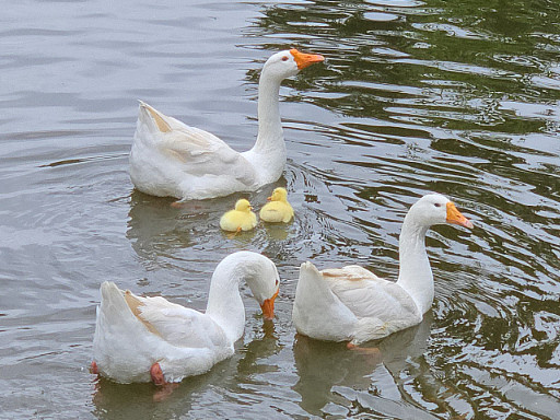
< A peaceful moment for the duck family. The baby goslings naturally followed the mother duck. >
It took ten months for this family to return. From abandonment and injury to healing, birth, and unexpected bonds, this was more than a story of survival. It was a journey of transformation. The duck family’s ten-month saga is a quiet miracle—written in small moments of crisis, care, and connection—and a lasting memory on the KAIST campus.
< The resident goose flock at KAIST’s pond naturally accepted the returning duck and goslings as part of their group. >
2025.06.10 View 6570
A 10-Month Journey of Tiny Flaps Completed: A Special Family Returns to KAIST Duck Pond
On the morning of June 9, 2025, gentle activity stirred early around the KAIST campus duck pond. It was the day a special family of ducks—and two goslings—were to be released back into the pond after spending a month in a temporary shelter. One by one, the ducklings cautiously emerged from their box, waddling toward the water's edge and scanning their surroundings, followed closely by their mother.
< The landscape manager from the KAIST Facilities Team releases the ducks and goslings. >
The mother duck, once a rescued loner who couldn’t integrate with the flock, returned triumphantly as the head of a new family—caring for both ducklings and goslings. Students and faculty looked on quietly, welcoming them back and reflecting on their remarkable 10-month journey.
The story began in July 2024, as a student filed a report of spotting two ducklings wandering near the pond without a mother. Based on their soft down, flat beaks, and lack of fear around humans, it was presumed they had been abandoned. Professor Won Do Heo of the Department of Biological Sciences—affectionately known as the “Goose Dad”—and the KAIST Facilities Team quickly stepped in to rescue them. After about a month of care, the ducklings were released back into the pond.
< On June 9, the day of the release, KAIST President Kwang-Hyung Lee (left), the former “Goose Dad,” and Professor Won Do Heo (right), the current “Goose Dad,” watched the flock as they freely wobbled about. >
At first, the ducklings seemed to adapt, but they started distancing themselves from the established goose flock. One eventually disappeared, and the remaining duckling was found injured by the pond during winter. Although KAIST typically avoids making human interference in the natural ecosystem, an exception was made to save the young duck’s life. It was put under the care of Professor Heo and the Facilities Team to regain its health within a month.
In the spring, the healed duck began laying eggs. Professor Heo supported the process by adjusting its diet, avoiding further intervention. On Children’s Day, May 5, the duck’s eggs hatched. The once-isolated duck had become a mother. Ten days later, on May 15, four goslings also hatched from the resident goose flock. With new life flourishing, the pond was more vibrant than ever.
< Rescued baby goslings near the pond, alongside the duck family that took them in. The mother duck—once a vulnerable duckling herself—had grown strong enough to care for others in need. >
But just days later, the mother goose disappeared, and two goslings—still unable to swim—were found shivering by the pond. Dahyeon Byeon, a student from Seoul National University who came for a visit on that day, reported this upon sighting, prompting another rescue. The vulnerable goslings were brought to the shelter to stay with the duck family.
Initially, the interspecies cohabitation was uneasy. But the mother duck did not reject the goslings. Slowly, they began to eat and sleep together, forming a new kind of family. After a month, they were released together into the pond—and to everyone’s surprise, the existing goose flock accepted both the goslings and the duck family.
< A peaceful moment for the duck family. The baby goslings naturally followed the mother duck. >
It took ten months for this family to return. From abandonment and injury to healing, birth, and unexpected bonds, this was more than a story of survival. It was a journey of transformation. The duck family’s ten-month saga is a quiet miracle—written in small moments of crisis, care, and connection—and a lasting memory on the KAIST campus.
< The resident goose flock at KAIST’s pond naturally accepted the returning duck and goslings as part of their group. >
2025.06.10 View 6570 -
 Life Springs at KAIST: A Tale of Two Special Campus Families
A Gift of Life on Teachers' Day: Baby Geese Born at KAIST Pond
On Teachers' Day, a meaningful miracle of life arrived at the KAIST campus. A pair of geese gave birth to two goslings by the duck pond.
< On Teachers' Day, a pair of geese and their goslings leisurely swim in the pond. >
The baby goslings, covered in yellow down, began exploring the pond's edge, scurrying about, while their aunt geese steadfastly stood by. Their curious glances, watchful gazes, playful hops on waterside rocks, and the procession of babies swimming behind their parents in the water melted the hearts of onlookers.
< As night falls on the duck pond, the goose family gathers among the reeds. >
This special new life, born on Teachers' Day, seems to symbolize the day's meaning of "care" and "growth." This wondrous scene of life brought warm comfort and joy to KAIST members, adding the inspiration of nature to a campus that is a space for research and learning.
< Under the protection of the adult geese, the goslings take their first steps, exploring the pond's grassy areas and rocks. >
This adorable family is already roaming the area leisurely, like the pond's owners. With the joy of life added to the spring-filled pond, warm smiles are spreading across the KAIST campus.
< The geese look around, surveying their surroundings, while caring for their goslings. >
The pond has now become a small but special haven for students and staff. This goose family, arriving on Teachers' Day, quietly reminds us of the meaning of care and learning conveyed by nature.
< The goose family shows care and growth, and warm moments together are anticipated. >
---
On Children's Day 2025, a Duck Becomes a Mother
In July 2024, a special guest arrived at the KAIST campus. With soft yellow down, waddling gait, and a flat beak, it was undeniably a baby duck. However, for some reason, its mother was nowhere to be seen. Given that it wasn't afraid of people and followed them well, it was clear that someone had abandoned the duck.
Fortunately, the baby duck was safely rescued thanks to prompt reporting by students.
< Two ducks found on a corner of campus, immediately after their rescue in summer 2024. >
The ducks, newly integrated into KAIST, seemed to adapt relatively peacefully to campus life. As new additions, they couldn't blend in with the existing goose flock that had settled on campus, but the geese didn't ostracize them either. Perhaps because they were awkward neighbors, there was hope that the ducks would soon join the existing goose flock.
< Following their rescue based on a student's report in summer 2024, the ducks adapted to campus life under the protection of the campus facility team and Professor Won Do Heo. >
Professor Won Do Heo of the Department of Biological Sciences, widely known as "Goose Dad," stepped forward to protect them along with the KAIST facility team. Professor Heo is well-known for consistently observing and protecting the campus geese and ducks, which are practically symbols of KAIST. Thanks to the care of the staff and Professor Heo, the two ducks were safely released back onto campus approximately one month after their rescue.
< A moment on campus: Before winter, the ducks lived separately from the goose flock, maintaining a certain distance. While there were no conflicts, they rarely socialized. >
However, as winter passed, sad news arrived. One duck went missing, and the remaining one was found injured by the pond. While the policy of the facility team and Professor Heo was to minimize intervention to allow campus animals to maintain their natural state, saving the injured duck was the top priority. After being isolated again for a month of recovery, the duck fully recovered and was able to greet spring under the sun.
< The mother duck left alone in winter: One went missing, and the remaining one was found injured. After indoor isolation and recovery, she was released back onto campus in the spring. >
As spring, the ducks' breeding season, began, Professor Heo decided to offer a little more help. When signs of egg-laying appeared, he consistently provided "special meals for pregnant mothers" throughout March. On the morning of May 5th, Children's Day, 28 days after the mother duck began incubating her eggs with the care and attention of KAIST members, new life finally hatched. It was a precious outcome achieved solely by the duck that had survived abandonment and injury, with no special protection other than food.
The duck, having overcome hardship and injury to stand alone, has now formed a new family. Although there is still some distance from the existing goose flock, it is expected that they will naturally find their place in the campus ecosystem, as KAIST's geese are not aggressive or exclusive. The KAIST goose flock already has experience protecting and raising five ducklings.
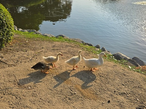
< A new beginning by the pond on Children's Day: On the morning of May 5th, the 28th day of incubation, four ducklings hatched by the pond. This was a natural hatching, achieved without protective equipment. >
A single duck brought a special spring to the KAIST campus on Children's Day. The outcome achieved by that small life, leading to the birth of a new family, also symbolizes the harmonious coexistence of people and animals on the KAIST campus. The careful intervention of KAIST members, providing only the necessary assistance from rescue to hatching, makes us reconsider what "desirable coexistence between animals and people" truly means.
2025.05.21 View 8614
Life Springs at KAIST: A Tale of Two Special Campus Families
A Gift of Life on Teachers' Day: Baby Geese Born at KAIST Pond
On Teachers' Day, a meaningful miracle of life arrived at the KAIST campus. A pair of geese gave birth to two goslings by the duck pond.
< On Teachers' Day, a pair of geese and their goslings leisurely swim in the pond. >
The baby goslings, covered in yellow down, began exploring the pond's edge, scurrying about, while their aunt geese steadfastly stood by. Their curious glances, watchful gazes, playful hops on waterside rocks, and the procession of babies swimming behind their parents in the water melted the hearts of onlookers.
< As night falls on the duck pond, the goose family gathers among the reeds. >
This special new life, born on Teachers' Day, seems to symbolize the day's meaning of "care" and "growth." This wondrous scene of life brought warm comfort and joy to KAIST members, adding the inspiration of nature to a campus that is a space for research and learning.
< Under the protection of the adult geese, the goslings take their first steps, exploring the pond's grassy areas and rocks. >
This adorable family is already roaming the area leisurely, like the pond's owners. With the joy of life added to the spring-filled pond, warm smiles are spreading across the KAIST campus.
< The geese look around, surveying their surroundings, while caring for their goslings. >
The pond has now become a small but special haven for students and staff. This goose family, arriving on Teachers' Day, quietly reminds us of the meaning of care and learning conveyed by nature.
< The goose family shows care and growth, and warm moments together are anticipated. >
---
On Children's Day 2025, a Duck Becomes a Mother
In July 2024, a special guest arrived at the KAIST campus. With soft yellow down, waddling gait, and a flat beak, it was undeniably a baby duck. However, for some reason, its mother was nowhere to be seen. Given that it wasn't afraid of people and followed them well, it was clear that someone had abandoned the duck.
Fortunately, the baby duck was safely rescued thanks to prompt reporting by students.
< Two ducks found on a corner of campus, immediately after their rescue in summer 2024. >
The ducks, newly integrated into KAIST, seemed to adapt relatively peacefully to campus life. As new additions, they couldn't blend in with the existing goose flock that had settled on campus, but the geese didn't ostracize them either. Perhaps because they were awkward neighbors, there was hope that the ducks would soon join the existing goose flock.
< Following their rescue based on a student's report in summer 2024, the ducks adapted to campus life under the protection of the campus facility team and Professor Won Do Heo. >
Professor Won Do Heo of the Department of Biological Sciences, widely known as "Goose Dad," stepped forward to protect them along with the KAIST facility team. Professor Heo is well-known for consistently observing and protecting the campus geese and ducks, which are practically symbols of KAIST. Thanks to the care of the staff and Professor Heo, the two ducks were safely released back onto campus approximately one month after their rescue.
< A moment on campus: Before winter, the ducks lived separately from the goose flock, maintaining a certain distance. While there were no conflicts, they rarely socialized. >
However, as winter passed, sad news arrived. One duck went missing, and the remaining one was found injured by the pond. While the policy of the facility team and Professor Heo was to minimize intervention to allow campus animals to maintain their natural state, saving the injured duck was the top priority. After being isolated again for a month of recovery, the duck fully recovered and was able to greet spring under the sun.
< The mother duck left alone in winter: One went missing, and the remaining one was found injured. After indoor isolation and recovery, she was released back onto campus in the spring. >
As spring, the ducks' breeding season, began, Professor Heo decided to offer a little more help. When signs of egg-laying appeared, he consistently provided "special meals for pregnant mothers" throughout March. On the morning of May 5th, Children's Day, 28 days after the mother duck began incubating her eggs with the care and attention of KAIST members, new life finally hatched. It was a precious outcome achieved solely by the duck that had survived abandonment and injury, with no special protection other than food.
The duck, having overcome hardship and injury to stand alone, has now formed a new family. Although there is still some distance from the existing goose flock, it is expected that they will naturally find their place in the campus ecosystem, as KAIST's geese are not aggressive or exclusive. The KAIST goose flock already has experience protecting and raising five ducklings.
< A new beginning by the pond on Children's Day: On the morning of May 5th, the 28th day of incubation, four ducklings hatched by the pond. This was a natural hatching, achieved without protective equipment. >
A single duck brought a special spring to the KAIST campus on Children's Day. The outcome achieved by that small life, leading to the birth of a new family, also symbolizes the harmonious coexistence of people and animals on the KAIST campus. The careful intervention of KAIST members, providing only the necessary assistance from rescue to hatching, makes us reconsider what "desirable coexistence between animals and people" truly means.
2025.05.21 View 8614 -
 A KAIST Research Team Observes the Processes of Memory and Cognition in Real Time
The human brain contains approximately 86 billion neurons and 600 trillion synapses that exchange signals between the neurons to help us control the various functions of the brain including cognition, emotion, and memory. Interestingly, the number of synapses decrease with age or as a result of diseases like Alzheimer’s, and research on synapses thus attracts a lot of attention. However, limitations have existed in observing the dynamics of synapse structures in real time.
On January 9, a joint research team led by Professor Won Do Heo from the KAIST Department of Biological Sciences, Professor Hyung-Bae Kwon from Johns Hopkins School of Medicine, and Professor Sangkyu Lee from the Institute for Basic Science (IBS) revealed that they have developed the world’s first technique to allow a real-time observation of synapse formation, extinction, and alterations.
Professor Heo’s team conjugated dimerization-dependent fluorescent proteins (ddFP) to synapses in order to observe the process in which synapses create connections between neurons in real time. The team named this technique SynapShot, by combining the words ‘synapse’ and snapshot’, and successfully tracked and observed the live formation and extinction processes of synapses as well as their dynamic changes.
< Figure 1. To observe dynamically changing synapses, dimerization-dependent fluorescent protein (ddFP) was expressed to observe flourescent signals upon synapse formation as ddFP enables fluorescence detection through reversible binding to pre- and postsynaptic terminals. >
Through a joint research project, the teams led by Professor Heo and Professor Sangkyu Lee at IBS together designed a SynapShot with green and red fluorescence, and were able to easily distinguish the synapse connecting two different neurons. Additionally, by combining an optogenetic technique that can control the function of a molecule using light, the team was able to observe the changes in the synapses while simultaneously inducing certain functions of the neurons using light.
Through more joint research with the team led by Professor Hyung-Bae Kwon at the Johns Hopkins School of Medicine, Professor Heo’s team induced several situations on live mice, including visual discrimination training, exercise, and anaesthesia, and used SynapShot to observe the changes in the synapses during each situation in real time. The observations revealed that each synapse could change fairly quickly and dynamically. This was the first-ever case in which the changes in synapses were observed in a live mammal.
< Figure 2. Microscopic photos observed through changes of the flourescence of the synapse sensor (SynapShot) by cultivating the neurons of an experimental rat and expressing the SynapShot. The changes in the synapse that is created when the pre- and post-synaptic terminals come into contact and the synapse that disappears after a certain period of time are measured by the fluorescence of the SynapShot. >
Professor Heo said, “Our group developed SynapShot through a collaboration with domestic and international research teams, and have opened up the possibility for first-hand live observations of the quick and dynamic changes of synapses, which was previously difficult to do. We expect this technique to revolutionize research methodology in the neurological field, and play an important role in brightening the future of brain science.”
This research, conducted by co-first authors Seungkyu Son (Ph.D. candidate), Jinsu Lee (Ph.D. candidate) and Dr. Kanghoon Jung from Johns Hopkins, was published in the online edition of Nature Methods on January 8 under the title “Real-time visualization of structural dynamics of synapses in live cells in vivo”, and will be printed in the February volume.
< Figure 3. Simultaneous use of green-SynapShot and red-SynapShot to distinguish and observe synapses with one post-terminal and different pre-terminals. >
< Figure 4. Dimer-dependent fluorescent protein (ddFP) exists as a green fluorescent protein as well as a red fluorescent protein, and can be applied together with blue light-activated optogenetic technology. After activating Tropomyosin receptor kinase B (TrkB) by blue light using optogenetic technology, the strengthening of synaptic connections through signals of brain-derived neurotrophic factor is observed using red-SynapShot. >
< Figure 5. Micrographs showing real-time changing synapses in the visual cortex of mice trained through visual training using in vivo imaging techniques such as two-photon microscopy as well as at the cellular level. >
This research was supported by Mid-Sized Research Funds and the Singularity Project from KAIST, and by IBS.
2024.01.18 View 12528
A KAIST Research Team Observes the Processes of Memory and Cognition in Real Time
The human brain contains approximately 86 billion neurons and 600 trillion synapses that exchange signals between the neurons to help us control the various functions of the brain including cognition, emotion, and memory. Interestingly, the number of synapses decrease with age or as a result of diseases like Alzheimer’s, and research on synapses thus attracts a lot of attention. However, limitations have existed in observing the dynamics of synapse structures in real time.
On January 9, a joint research team led by Professor Won Do Heo from the KAIST Department of Biological Sciences, Professor Hyung-Bae Kwon from Johns Hopkins School of Medicine, and Professor Sangkyu Lee from the Institute for Basic Science (IBS) revealed that they have developed the world’s first technique to allow a real-time observation of synapse formation, extinction, and alterations.
Professor Heo’s team conjugated dimerization-dependent fluorescent proteins (ddFP) to synapses in order to observe the process in which synapses create connections between neurons in real time. The team named this technique SynapShot, by combining the words ‘synapse’ and snapshot’, and successfully tracked and observed the live formation and extinction processes of synapses as well as their dynamic changes.
< Figure 1. To observe dynamically changing synapses, dimerization-dependent fluorescent protein (ddFP) was expressed to observe flourescent signals upon synapse formation as ddFP enables fluorescence detection through reversible binding to pre- and postsynaptic terminals. >
Through a joint research project, the teams led by Professor Heo and Professor Sangkyu Lee at IBS together designed a SynapShot with green and red fluorescence, and were able to easily distinguish the synapse connecting two different neurons. Additionally, by combining an optogenetic technique that can control the function of a molecule using light, the team was able to observe the changes in the synapses while simultaneously inducing certain functions of the neurons using light.
Through more joint research with the team led by Professor Hyung-Bae Kwon at the Johns Hopkins School of Medicine, Professor Heo’s team induced several situations on live mice, including visual discrimination training, exercise, and anaesthesia, and used SynapShot to observe the changes in the synapses during each situation in real time. The observations revealed that each synapse could change fairly quickly and dynamically. This was the first-ever case in which the changes in synapses were observed in a live mammal.
< Figure 2. Microscopic photos observed through changes of the flourescence of the synapse sensor (SynapShot) by cultivating the neurons of an experimental rat and expressing the SynapShot. The changes in the synapse that is created when the pre- and post-synaptic terminals come into contact and the synapse that disappears after a certain period of time are measured by the fluorescence of the SynapShot. >
Professor Heo said, “Our group developed SynapShot through a collaboration with domestic and international research teams, and have opened up the possibility for first-hand live observations of the quick and dynamic changes of synapses, which was previously difficult to do. We expect this technique to revolutionize research methodology in the neurological field, and play an important role in brightening the future of brain science.”
This research, conducted by co-first authors Seungkyu Son (Ph.D. candidate), Jinsu Lee (Ph.D. candidate) and Dr. Kanghoon Jung from Johns Hopkins, was published in the online edition of Nature Methods on January 8 under the title “Real-time visualization of structural dynamics of synapses in live cells in vivo”, and will be printed in the February volume.
< Figure 3. Simultaneous use of green-SynapShot and red-SynapShot to distinguish and observe synapses with one post-terminal and different pre-terminals. >
< Figure 4. Dimer-dependent fluorescent protein (ddFP) exists as a green fluorescent protein as well as a red fluorescent protein, and can be applied together with blue light-activated optogenetic technology. After activating Tropomyosin receptor kinase B (TrkB) by blue light using optogenetic technology, the strengthening of synaptic connections through signals of brain-derived neurotrophic factor is observed using red-SynapShot. >
< Figure 5. Micrographs showing real-time changing synapses in the visual cortex of mice trained through visual training using in vivo imaging techniques such as two-photon microscopy as well as at the cellular level. >
This research was supported by Mid-Sized Research Funds and the Singularity Project from KAIST, and by IBS.
2024.01.18 View 12528 -
 Professor Won Do Heo Receives 'Scientist of the Month Award'
Professor Won Do Heo of the Department of Biological Sciences was selected as the “Scientist of the Month” for April 2017 by the Ministry of Science, ICT and Future Planning and the National Research Foundation of Korea.
Professor Heo was recognized for his suggestion of a new biological research method developing various optogenetics technology which controls cell function by using light. He developed the technology using lasers or LED light, without the need for surgery or drug administration, to identify the cause of diseases related to calcium ions such as Alzheimer’s disease and cancer.
The general technique used in optogenetics, that control cells in the body with light, is the simple activation and deactivation of neurons.
Professor Heo developed a calcium ion channel activation technique (OptoSTIM1) to activate calcium ions in the body using light. He also succeeded in increasing calcium concentrations with light to enhance the memory capacity of mice two-fold.
Using this technology, the desired amount and residing time of calcium ion influx can be controlled by changing light intensity and exposure periods, enabling the function of a single cell or various cells in animal tissue to be controlled remotely.
The experimental results showed that calcium ion influx can be activated in cells that are affected by calcium ions, such as normal cells, cancer cells, and human embryonic stem cells. By controlling calcium concentrations with light, it is possible to control biological phenomena, such as cellular growth, neurotransmitter transmission, muscle contraction, and hormone control.
Professor Heo said, “Until now, it was standard to use optogenetics to activate neurons using channelrhodopsin. The development of this new optogenetic technique using calcium ion channel activation can be applied to various biological studies, as well as become an essential research technique in neurobiology.
The “Scientist of the Month Award” is given every month to one researcher who made significant contributions to the advancement of science and technology with their outstanding research achievement. The awardee will receive prize money of ten million won.
2017.04.07 View 13613
Professor Won Do Heo Receives 'Scientist of the Month Award'
Professor Won Do Heo of the Department of Biological Sciences was selected as the “Scientist of the Month” for April 2017 by the Ministry of Science, ICT and Future Planning and the National Research Foundation of Korea.
Professor Heo was recognized for his suggestion of a new biological research method developing various optogenetics technology which controls cell function by using light. He developed the technology using lasers or LED light, without the need for surgery or drug administration, to identify the cause of diseases related to calcium ions such as Alzheimer’s disease and cancer.
The general technique used in optogenetics, that control cells in the body with light, is the simple activation and deactivation of neurons.
Professor Heo developed a calcium ion channel activation technique (OptoSTIM1) to activate calcium ions in the body using light. He also succeeded in increasing calcium concentrations with light to enhance the memory capacity of mice two-fold.
Using this technology, the desired amount and residing time of calcium ion influx can be controlled by changing light intensity and exposure periods, enabling the function of a single cell or various cells in animal tissue to be controlled remotely.
The experimental results showed that calcium ion influx can be activated in cells that are affected by calcium ions, such as normal cells, cancer cells, and human embryonic stem cells. By controlling calcium concentrations with light, it is possible to control biological phenomena, such as cellular growth, neurotransmitter transmission, muscle contraction, and hormone control.
Professor Heo said, “Until now, it was standard to use optogenetics to activate neurons using channelrhodopsin. The development of this new optogenetic technique using calcium ion channel activation can be applied to various biological studies, as well as become an essential research technique in neurobiology.
The “Scientist of the Month Award” is given every month to one researcher who made significant contributions to the advancement of science and technology with their outstanding research achievement. The awardee will receive prize money of ten million won.
2017.04.07 View 13613