Biomaterials+Engineering
-
 Cooperative Tumor Cell Membrane-Targeted Phototherapy
A KAIST research team led by Professor Ji-Ho Park in the Bio and Brain Engineering Department at KAIST developed a technology for the effective treatment of cancer by delivering synthetic receptors throughout tumor tissue. The study, led by Ph.D. candidate Heegon Kim, was published online in Nature Communications on June 19.
Cancer targeted therapy generally refers to therapy targeting specific molecules that are involved in the growth and generation of cancer. The targeted delivery of therapeutics using targeting agents such as antibodies or nanomaterials has improved the precision and safety of cancer therapy.
However, the paucity and heterogeneity of identified molecular targets within tumors have resulted in poor and uneven distribution of targeted agents, thus compromising treatment outcomes.
To solve this problem, the team constructed a cooperative targeting system in which synthetic and biological nanocomponents participate together in the tumor cell membrane-selective localization of synthetic receptors to amplify the subsequent targeting of therapeutics. Here, synthetic and biological nanocomponents refer to liposomes and extracellular vesicles, respectively.
The synthetic receptors are first delivered selectively to tumor cell membranes in the perivascular region using liposomes. By hitchhiking with extracellular vesicles secreted by the cells, the synthetic receptors are transferred to neighboring cells and further spread throughout the tumor tissues where the molecular targets are limited.
Hitchhiking extracellular vesicles for delivery of synthetic receptors was possible since extracellular vesicles, such as exosomes, mediate intercellular communications by transferring various biological components such as lipids, cytosolic proteins, and RNA through a membrane fusion process. They also play a supportive role in promoting tumor progression in that tumor-derived extracellular vesicles deliver oncogenic signals to normal host cells.
The team showed that this tumor cell membrane-targeted delivery of synthetic receptors led to a uniform distribution of synthetic receptors throughout a tumor and subsequently led to enhanced phototherapeutic efficacy of the targeted photosensitizer.
Professor Park said, “The cooperative tumor targeting system is expected to be applied in treating various diseases that are hard to target.”
The research was funded by the Basic Science Research Program through the National Research Foundation funded by the Ministry of Science, ICT & Future Planning, and the National R&D Program for Cancer Control funded by the Ministry for Health and Welfare.
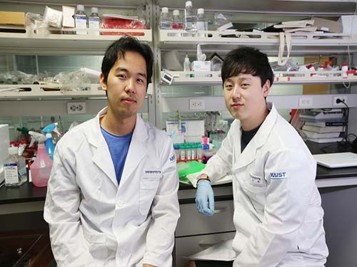
(Ph.D. candidates Hee Gon Kim (left) and Chanhee Oh)
Figure 1. A schematic of a cooperative tumor targeting system via delivery of synthetic receptors.
Figure 2. A confocal microscopic image of a tumor section after cooperative targeting by synthetic receptor delivery. Green and magenta represent vessels and therapeutic agents inside a tumor respectively.
2017.07.07 View 12042
 -
 Seeing Inside Cells with Fiber Optics
Professor Jiho Park’s research team was successful in receiving minute optical signals from inside the cell using optical nano fibers.
Through the invention of this technology, we can now look inside cells in high resolution without the use of equipment such as endoscopes that damage cells. We will be able to study the biological phenomena within cells, and thus cure diseases more effectively.
Recently, ultra high resolution microscopes have been used to analyze incubated cells. However, because of the need for a very complex and large system, it had been impossible to monitor cells in the less transparent areas of the body in real time.
The research team created the wire with a semiconductor created with tin oxides to be only 100 nanometers in diameter (1nanometer= 1/1billion meters).
The nanowire is connected to the end of the optical fiber, and the light that comes through the optical fiber is transmitted to particular spots in the cell, and the optical signals from the cell are retrieved back from the cell as well
Together with this, based on the fact that nanowires do not damage cells, the research team covered the end of the wire with a photo reactive material and entered this into the cell. They were able to check that the material reacted to light and entered the cell when they transmitted light
Accordingly, this showed the possibilities of the use of this technology as a method of treatment to effectively transfer the medication into the cells.
Prof. Jiho Park stated that “in this research, we only used cells incubated outside the human body, but soon we will use this technology to stimulate and control cells within the body in a minute scale” as well as that “soon, we will be able to study the biological phenomena inside a cell to study diseases and apply this to cure them more effectively”.
This research result has been published in the online publication of ‘Nature Nanotechnology’ on December 18.
This study was done through the cooperation of various schools. Besides Prof. Jiho Park, Prof. Seungman Yang from the Biochemistry department, and Doctor Chuljoon Huh from KAIST, Prof. Yeonho Choi from Biomedical Science department of Korea University, Professor Peidon Yang and Doctor Ruoxue Yan from UC Berkeley’s chemistry department, and Luke Lee from UC Berkeley’s bioengineering department participated in the project.
2012.01.31 View 10800