%70%72%6f%74%65%69%6e
-
 InnoCORE Research Group Successfully Achieves AI Protein Design with Nobel Laureate David Baker
< (From left) Professor Gyu Rie Lee, Professor David Baker >
Under the foundation of research cooperation established through the Ministry of Science and ICT's InnoCORE (InnoCORE) project, KAIST InnoCORE researchers have derived meaningful research results. Following a visit by Professor David Baker (University of Washington, USA), the 2024 Nobel Laureate in Chemistry, KAIST has revealed research findings on designing proteins that accurately recognize desired compounds using AI through joint research.
KAIST announced on April 9th that Professor Gyu Rie Lee of the Department of Biological Sciences—a researcher participating in the AI-CRED Innovative Drug InnoCORE Research Group—successfully designed artificial proteins that selectively recognize specific compounds using AI through joint research with Professor David Baker.
This research is characterized by using AI to design proteins that recognize specific compounds from scratch (de novo) and implementing them as functional biosensors. While the conventional approach mainly involved searching natural proteins or modifying some of their functions, this research is highly significant in that it ‘custom-built’ proteins with desired functions through AI-based design and even completed experimental verification.
In particular, the research team successfully designed a protein that selectively recognizes the stress hormone cortisol and implemented an AI-designed biosensor based on it. This is evaluated as a case that extends beyond protein design to actual measurable sensor technology, solving the long-standing challenge of small-molecule recognition in the field of protein design.
These research results are expected to be utilized in various fields such as disease diagnosis, new drug development, and environmental monitoring in the future. It can precisely detect biomarkers in the blood to diagnose diseases early and contribute to the development of targeted therapies through the design of proteins that selectively recognize specific molecules. Furthermore, it is expected that the implementation of customized biosensor technology will become possible, such as real-time monitoring of air and water quality through the development of sensors that detect environmental pollutants.
Designing new proteins (de novo proteins) that recognize compounds has been considered a challenge in the field of protein design for a long time because it requires precise calculations at the atomic level. The research team developed an AI model that precisely reflects protein-ligand interactions and successfully designed binding proteins using it.
As a result, artificial binding proteins were designed for six types of compounds, including metabolites and small-molecule drugs, and their functions were verified through experiments. In particular, a cortisol biosensor was developed by designing a chemical-induced dimer based on a new protein that binds with cortisol. A provisional patent for the relevant design technology has been filed in the United States.
Professor Gyu Rie Lee stated, “This research experimentally proves that AI can be used to design proteins that precisely recognize specific compounds,” and added, “We plan to expand this into protein design technology that can be utilized in various fields such as disease diagnosis, new drug development, and environmental monitoring in the future.”
Professor Gyu Rie Lee of the KAIST Department of Biological Sciences participated in this research as the first author, and Professor David Baker as the corresponding author. The study was published in the international academic journal Nature Communications on March 28, 2026. ※ Paper Title: Small-molecule binding and sensing with a designed protein family DOI: https://doi.org/10.1038/s41467-026-70953-8 Authors: Gyu Rie Lee, Samuel J. Pellock, Christoffer Norn, Doug Tischer, Justas Dauparas, Ivan Anishchenko, Jaron A. M. Mercer, Alex Kang, Asim K. Bera, Hannah Nguyen, Evans Brackenbrough, Banumathi Sankaran, Inna Goreshnik, Dionne Vafeados, Nicole Roullier, Hannah L. Han, Brian Coventry, Hugh K. Haddox, David R. Liu, Andy Hsien-Wei Yeh & David Baker
< Image of Research Content Summary >
Professor Gyu Rie Lee is a new professor who joined KAIST in February 2025 and leads the Protein Design Laboratory. She possesses world-class expertise in the field of precise protein complex design at the atomic level and is performing various research projects such as AI-based protein design, artificial enzyme design, and RNA-recognizing protein development. She is also participating as a mentor professor in the AI-CRED Innovative Drug Research Group of the InnoCORE project, conducting research on enzyme and peptide drug design.
Professor Lee conducted research as a postdoctoral researcher and Staff Scientist in Professor David Baker’s laboratory (University of Washington, USA, Howard Hughes Medical Institute) from 2018 to 2024. Professor David Baker is a world-renowned scholar in the field of protein structure prediction and design and was awarded the Nobel Prize in Chemistry in 2024.
Director Do-Heon Lee, a mentor professor of the AI-CRED Innovative Drug Research Group, stated, “This achievement is a meaningful result derived through cooperation between InnoCORE researchers and a global scholar,” and added, “We will further strengthen our research capabilities based on active research collaboration with postdoctoral researchers recruited through the InnoCORE project to continue creating innovative results in the AI drug development and bio-fields.”
Meanwhile, KAIST will host a lecture on Thursday, April 9th at 4 PM in the KI Building Fusion Hall featuring Professor David Baker and Professor Hannele Ruohola-Baker (University of Washington, USA) under the theme of ‘Advances in AI-powered protein design and biomedical science’ to mark Professor David Baker’s visit to Korea. This event is held with the support of the KAIST International Scholar Invitation Program, KAI-X, the InnoCORE AI-CRED Innovative Drug Group, and the Ministry of Science and ICT’s Overseas Excellent Research Institute Cooperation Hub Construction Project.
< Poster for Professor David Baker’s Invited Lecture >
KAIST President Kwang Hyung Lee stated, “Through cooperation with Nobel Laureate Professor David Baker, we have derived a meaningful achievement in AI-based protein design,” and added, “This research is an example showing that KAIST is leading innovative research alongside world-class research institutions.”
Meanwhile, the KAIST InnoCORE Research Group aims to accelerate AI-based scientific and technological innovation and promote global joint research by supporting top-tier domestic and international postdoctoral researchers to devote themselves to the development of AI convergence technology in a cutting-edge collective research environment. As the lead institution, KAIST operates the ▲Hyper-scale Large Language Model Innovation Research Group ▲AI-based Intelligent Design-Manufacturing Integration Research Group ▲AI-CRED Innovative Drug Research Group and ▲AI-Transformed Aerospace Research Group.
2026.04.09 View 1445
InnoCORE Research Group Successfully Achieves AI Protein Design with Nobel Laureate David Baker
< (From left) Professor Gyu Rie Lee, Professor David Baker >
Under the foundation of research cooperation established through the Ministry of Science and ICT's InnoCORE (InnoCORE) project, KAIST InnoCORE researchers have derived meaningful research results. Following a visit by Professor David Baker (University of Washington, USA), the 2024 Nobel Laureate in Chemistry, KAIST has revealed research findings on designing proteins that accurately recognize desired compounds using AI through joint research.
KAIST announced on April 9th that Professor Gyu Rie Lee of the Department of Biological Sciences—a researcher participating in the AI-CRED Innovative Drug InnoCORE Research Group—successfully designed artificial proteins that selectively recognize specific compounds using AI through joint research with Professor David Baker.
This research is characterized by using AI to design proteins that recognize specific compounds from scratch (de novo) and implementing them as functional biosensors. While the conventional approach mainly involved searching natural proteins or modifying some of their functions, this research is highly significant in that it ‘custom-built’ proteins with desired functions through AI-based design and even completed experimental verification.
In particular, the research team successfully designed a protein that selectively recognizes the stress hormone cortisol and implemented an AI-designed biosensor based on it. This is evaluated as a case that extends beyond protein design to actual measurable sensor technology, solving the long-standing challenge of small-molecule recognition in the field of protein design.
These research results are expected to be utilized in various fields such as disease diagnosis, new drug development, and environmental monitoring in the future. It can precisely detect biomarkers in the blood to diagnose diseases early and contribute to the development of targeted therapies through the design of proteins that selectively recognize specific molecules. Furthermore, it is expected that the implementation of customized biosensor technology will become possible, such as real-time monitoring of air and water quality through the development of sensors that detect environmental pollutants.
Designing new proteins (de novo proteins) that recognize compounds has been considered a challenge in the field of protein design for a long time because it requires precise calculations at the atomic level. The research team developed an AI model that precisely reflects protein-ligand interactions and successfully designed binding proteins using it.
As a result, artificial binding proteins were designed for six types of compounds, including metabolites and small-molecule drugs, and their functions were verified through experiments. In particular, a cortisol biosensor was developed by designing a chemical-induced dimer based on a new protein that binds with cortisol. A provisional patent for the relevant design technology has been filed in the United States.
Professor Gyu Rie Lee stated, “This research experimentally proves that AI can be used to design proteins that precisely recognize specific compounds,” and added, “We plan to expand this into protein design technology that can be utilized in various fields such as disease diagnosis, new drug development, and environmental monitoring in the future.”
Professor Gyu Rie Lee of the KAIST Department of Biological Sciences participated in this research as the first author, and Professor David Baker as the corresponding author. The study was published in the international academic journal Nature Communications on March 28, 2026. ※ Paper Title: Small-molecule binding and sensing with a designed protein family DOI: https://doi.org/10.1038/s41467-026-70953-8 Authors: Gyu Rie Lee, Samuel J. Pellock, Christoffer Norn, Doug Tischer, Justas Dauparas, Ivan Anishchenko, Jaron A. M. Mercer, Alex Kang, Asim K. Bera, Hannah Nguyen, Evans Brackenbrough, Banumathi Sankaran, Inna Goreshnik, Dionne Vafeados, Nicole Roullier, Hannah L. Han, Brian Coventry, Hugh K. Haddox, David R. Liu, Andy Hsien-Wei Yeh & David Baker
< Image of Research Content Summary >
Professor Gyu Rie Lee is a new professor who joined KAIST in February 2025 and leads the Protein Design Laboratory. She possesses world-class expertise in the field of precise protein complex design at the atomic level and is performing various research projects such as AI-based protein design, artificial enzyme design, and RNA-recognizing protein development. She is also participating as a mentor professor in the AI-CRED Innovative Drug Research Group of the InnoCORE project, conducting research on enzyme and peptide drug design.
Professor Lee conducted research as a postdoctoral researcher and Staff Scientist in Professor David Baker’s laboratory (University of Washington, USA, Howard Hughes Medical Institute) from 2018 to 2024. Professor David Baker is a world-renowned scholar in the field of protein structure prediction and design and was awarded the Nobel Prize in Chemistry in 2024.
Director Do-Heon Lee, a mentor professor of the AI-CRED Innovative Drug Research Group, stated, “This achievement is a meaningful result derived through cooperation between InnoCORE researchers and a global scholar,” and added, “We will further strengthen our research capabilities based on active research collaboration with postdoctoral researchers recruited through the InnoCORE project to continue creating innovative results in the AI drug development and bio-fields.”
Meanwhile, KAIST will host a lecture on Thursday, April 9th at 4 PM in the KI Building Fusion Hall featuring Professor David Baker and Professor Hannele Ruohola-Baker (University of Washington, USA) under the theme of ‘Advances in AI-powered protein design and biomedical science’ to mark Professor David Baker’s visit to Korea. This event is held with the support of the KAIST International Scholar Invitation Program, KAI-X, the InnoCORE AI-CRED Innovative Drug Group, and the Ministry of Science and ICT’s Overseas Excellent Research Institute Cooperation Hub Construction Project.
< Poster for Professor David Baker’s Invited Lecture >
KAIST President Kwang Hyung Lee stated, “Through cooperation with Nobel Laureate Professor David Baker, we have derived a meaningful achievement in AI-based protein design,” and added, “This research is an example showing that KAIST is leading innovative research alongside world-class research institutions.”
Meanwhile, the KAIST InnoCORE Research Group aims to accelerate AI-based scientific and technological innovation and promote global joint research by supporting top-tier domestic and international postdoctoral researchers to devote themselves to the development of AI convergence technology in a cutting-edge collective research environment. As the lead institution, KAIST operates the ▲Hyper-scale Large Language Model Innovation Research Group ▲AI-based Intelligent Design-Manufacturing Integration Research Group ▲AI-CRED Innovative Drug Research Group and ▲AI-Transformed Aerospace Research Group.
2026.04.09 View 1445 -
 Longevity mediated by suppressing age-associated circRNA
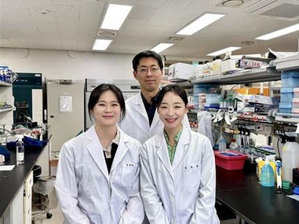
< (Back row from left) Prof. Yoon Ki Kim, Prof. Seung-Jae V. Lee, and Gwangrog Lee; (Front row from left) Dr. Sung Ho Boo, Sieun S. Kim, Seokjin Ham, and (top) Donghun Lee >
Cells in our bodies produce RNA based on genetic information stored in DNA, and RNA serves as a blueprint for making proteins. Researchers at our university have discovered a new phenomenon: removing 'circular RNA' that accumulates in cells as we age can slow down aging and extend lifespan. This study provides crucial clues for uncovering the principles of aging and developing treatment strategies for related diseases.
Professor Seung-Jae V. Lee’s research team (RNA-Mediated Healthspan and Longevity Research Center) from the Department of Biological Sciences, in collaboration with research teams led by Professors Yoon Ki Kim and Gwangrog Lee, announced on the 18th that they discovered the RNASEK protein—an enzyme that degrades circular RNA—plays a vital role in slowing aging and extending lifespan.
Until now, circular RNA has been regarded mainly as an aging marker because of its stability, which allows it to accumulate over time. However, the molecular mechanism for removing this RNA and its direct link to aging had not been clearly identified. The research team conducted this study to determine how the accumulation of circular RNA affects aging and whether an intracellular management system exists to regulate it.
Using Caenorhabditis elegans (C. elegans), a short-lived roundworm widely used in aging research, the team first confirmed that the circular RNA-degrading enzyme RNASEK is essential for longevity. They also discovered that as aging progresses, the amount of RNASEK decreases, resulting in an abnormal accumulation of circular RNA within cells.
Conversely, artificially increasing the levels of RNASEK (overexpression) extended the lifespan and allowed the organisms to survive longer in a healthy state. This implies that the process of appropriately removing cellular circular RNA is critical for maintaining health and longevity.
The research team also found that RNASEK prevents the toxic aggregation of circular RNAs in aged organisms. . When RNASEK is deficient and circular RNA accumulates, "stress granules" form abnormally inside the cell, which can impair cellular functions and accelerate aging.
RNASEK works alongside the chaperone protein HSP90 (which helps proteins avoid misfolding or clumping) to inhibit the formation of these stress granules and help cells maintain a normal state. Notably, this phenomenon was observed not only in C. elegans but also in human cells. In mammals, RNASEK also functions to directly degrade circular RNA; a deficiency of RNASEK in human cells and mouse models led to premature aging.
< Diagram showing progress toward longevity or aging depending on circular RNA and the removal enzyme RNASEK >
The researchers explained that this study is significant as it identifies a mechanism for regulating aging at the RNA level. They suggested that research using RNASEK to control circular RNA could lead to the development of treatment strategies for human aging and degenerative diseases.
Professor Seung-Jae V. Lee of KAIST, who led the study, explained, "Until now, circular RNA was merely regarded as a marker of aging that accumulates over time due to its stability. This study proves that circular RNA accumulated during aging actually induces aging, and that RNASEK, which removes it, is a key regulator that slows aging and induces healthy longevity."
< (AI-generated image) Longevity induced by the circular RNA-removing enzyme RNASEK >
Drs. Sieun S. Kim, Seokjin Ham, Sung Ho Boo, and Donghun Lee from the KAIST Department of Biological Sciences participated as joint first authors. The research results were published on February 24 in the world-renowned scientific journal Molecular Cell.
Paper Title: Ribonuclease $\kappa$ promotes longevity by preventing age-associated accumulation of circular RNA in stress granules
DOI: 10.1016/j.molcel.2026.01.031
This research was conducted with support from the Leader Researcher Program of the National Research Foundation of Korea.
2026.03.18 View 1677
Longevity mediated by suppressing age-associated circRNA
< (Back row from left) Prof. Yoon Ki Kim, Prof. Seung-Jae V. Lee, and Gwangrog Lee; (Front row from left) Dr. Sung Ho Boo, Sieun S. Kim, Seokjin Ham, and (top) Donghun Lee >
Cells in our bodies produce RNA based on genetic information stored in DNA, and RNA serves as a blueprint for making proteins. Researchers at our university have discovered a new phenomenon: removing 'circular RNA' that accumulates in cells as we age can slow down aging and extend lifespan. This study provides crucial clues for uncovering the principles of aging and developing treatment strategies for related diseases.
Professor Seung-Jae V. Lee’s research team (RNA-Mediated Healthspan and Longevity Research Center) from the Department of Biological Sciences, in collaboration with research teams led by Professors Yoon Ki Kim and Gwangrog Lee, announced on the 18th that they discovered the RNASEK protein—an enzyme that degrades circular RNA—plays a vital role in slowing aging and extending lifespan.
Until now, circular RNA has been regarded mainly as an aging marker because of its stability, which allows it to accumulate over time. However, the molecular mechanism for removing this RNA and its direct link to aging had not been clearly identified. The research team conducted this study to determine how the accumulation of circular RNA affects aging and whether an intracellular management system exists to regulate it.
Using Caenorhabditis elegans (C. elegans), a short-lived roundworm widely used in aging research, the team first confirmed that the circular RNA-degrading enzyme RNASEK is essential for longevity. They also discovered that as aging progresses, the amount of RNASEK decreases, resulting in an abnormal accumulation of circular RNA within cells.
Conversely, artificially increasing the levels of RNASEK (overexpression) extended the lifespan and allowed the organisms to survive longer in a healthy state. This implies that the process of appropriately removing cellular circular RNA is critical for maintaining health and longevity.
The research team also found that RNASEK prevents the toxic aggregation of circular RNAs in aged organisms. . When RNASEK is deficient and circular RNA accumulates, "stress granules" form abnormally inside the cell, which can impair cellular functions and accelerate aging.
RNASEK works alongside the chaperone protein HSP90 (which helps proteins avoid misfolding or clumping) to inhibit the formation of these stress granules and help cells maintain a normal state. Notably, this phenomenon was observed not only in C. elegans but also in human cells. In mammals, RNASEK also functions to directly degrade circular RNA; a deficiency of RNASEK in human cells and mouse models led to premature aging.
< Diagram showing progress toward longevity or aging depending on circular RNA and the removal enzyme RNASEK >
The researchers explained that this study is significant as it identifies a mechanism for regulating aging at the RNA level. They suggested that research using RNASEK to control circular RNA could lead to the development of treatment strategies for human aging and degenerative diseases.
Professor Seung-Jae V. Lee of KAIST, who led the study, explained, "Until now, circular RNA was merely regarded as a marker of aging that accumulates over time due to its stability. This study proves that circular RNA accumulated during aging actually induces aging, and that RNASEK, which removes it, is a key regulator that slows aging and induces healthy longevity."
< (AI-generated image) Longevity induced by the circular RNA-removing enzyme RNASEK >
Drs. Sieun S. Kim, Seokjin Ham, Sung Ho Boo, and Donghun Lee from the KAIST Department of Biological Sciences participated as joint first authors. The research results were published on February 24 in the world-renowned scientific journal Molecular Cell.
Paper Title: Ribonuclease $\kappa$ promotes longevity by preventing age-associated accumulation of circular RNA in stress granules
DOI: 10.1016/j.molcel.2026.01.031
This research was conducted with support from the Leader Researcher Program of the National Research Foundation of Korea.
2026.03.18 View 1677 -
 KAIST Surpasses the Limits of AlphaFold… AI Now Predicts Whether Drugs Actually Work
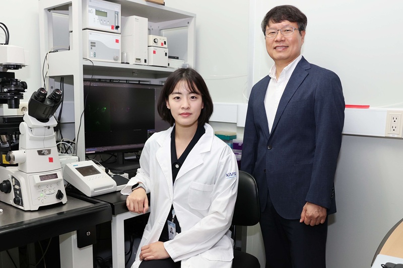
<(From Left) Ph.D candidate Hyojin Son, Professor Gwan-su Yi>
Proteins in our body function like switches. When a drug binds to a protein, the structure at the binding site changes, and this structural change propagates throughout the protein, turning its function on or off. Google DeepMind’s AlphaFold3 successfully predicted whether drugs bind to proteins and the three-dimensional structure of binding sites. However, it could not predict how signals propagate inside the protein after drug binding, how the entire structure changes, or whether the protein’s function is ultimately activated or inhibited. KAIST researchers have developed an AI that predicts not only whether a drug binds but whether it actually works.
KAIST (President Kwang Hyung Lee) announced on the 4th of March that a research team led by Professor Gwan-Su Yi of the Department of Bio and Brain Engineering has developed an artificial intelligence model called “GPCRact” that predicts whether candidate molecules not only bind to G-protein-coupled receptors (GPCRs)—a major drug target—but also actually activate the protein.
GPCRs act as “signal receivers” on the surface of cells. When hormones, neurotransmitters, or drugs send signals from outside the cell, GPCRs function as gates that receive these signals and transmit them into the cell. There are about 800 types of GPCRs in the human body, and roughly 30–40% of currently marketed drugs target them. They are key proteins involved in numerous physiological functions, including heart rate regulation, blood pressure control, pain sensing, immune responses, and emotional regulation.
However, a drug binding to a GPCR does not always trigger the desired biological function. Structural changes inside the protein and subsequent signal transmission determine whether the drug actually produces an effect. This process is known as allosteric signal propagation.
The research team designed the AI to learn the drug action process in two stages: ① the drug–target binding stage, and ② the intracellular signal propagation stage within the protein. The three-dimensional protein structure was represented as an atom-level graph, and an attention mechanism was applied to enable the model to learn important signaling pathways. Through this approach, the AI analyzes not only the drug binding signal but also the internal signaling pathways of the protein to predict whether the protein becomes activated.
As a result, the model significantly improved the prediction performance of drug activity even in proteins with complex structures that existing models struggled to analyze. Importantly, the model does not simply output “active” or “inactive.” It also presents the key internal signaling pathways that form the basis of its predictions, overcoming the limitations of so-called “black-box AI.”
<Schematic diagram of drug activity prediction and mechanism interpretation using the GPCRact artificial intelligence model>
This represents an important advance, as it allows researchers to interpret and verify predictions while simultaneously improving the reliability and efficiency of drug discovery. In the future, the model is expected to serve as a precision drug discovery AI platform capable of predicting not only whether drugs bind to GPCRs but also whether they truly activate them in various diseases targeting GPCRs.
<AI-generated image to help illustrate the research>
Professor Gwan-Su Yi explained, “Allosteric structural change refers to a phenomenon in which a drug binds to one part of a protein and its influence propagates internally, altering the function of other regions,” adding, “The key contribution of this research is incorporating this operational principle into deep learning.” He further noted, “We plan to expand the model to various proteins and ultimately develop technologies capable of predicting cellular and whole-body responses.”
Ph.D candidate Hyojin Son participated as the first author in this study. The paper was published on January 15 in the international journal Briefings in Bioinformatics, one of the leading journals in the field of bioinformatics.
※ Paper title: “GPCRact: a hierarchical framework for predicting ligand-induced GPCR activity via allosteric communication modeling”
DOI: https://doi.org/10.1093/bib/bbaf719※ Author information: Hyojin Son (KAIST, first author), Gwan-Su Yi (KAIST, corresponding author)
This research was supported by the Basic Research Program for Individual Research funded by the Ministry of Science and ICT and the National Research Foundation of Korea (RS-2025-24533057).
2026.03.09 View 1236
KAIST Surpasses the Limits of AlphaFold… AI Now Predicts Whether Drugs Actually Work
<(From Left) Ph.D candidate Hyojin Son, Professor Gwan-su Yi>
Proteins in our body function like switches. When a drug binds to a protein, the structure at the binding site changes, and this structural change propagates throughout the protein, turning its function on or off. Google DeepMind’s AlphaFold3 successfully predicted whether drugs bind to proteins and the three-dimensional structure of binding sites. However, it could not predict how signals propagate inside the protein after drug binding, how the entire structure changes, or whether the protein’s function is ultimately activated or inhibited. KAIST researchers have developed an AI that predicts not only whether a drug binds but whether it actually works.
KAIST (President Kwang Hyung Lee) announced on the 4th of March that a research team led by Professor Gwan-Su Yi of the Department of Bio and Brain Engineering has developed an artificial intelligence model called “GPCRact” that predicts whether candidate molecules not only bind to G-protein-coupled receptors (GPCRs)—a major drug target—but also actually activate the protein.
GPCRs act as “signal receivers” on the surface of cells. When hormones, neurotransmitters, or drugs send signals from outside the cell, GPCRs function as gates that receive these signals and transmit them into the cell. There are about 800 types of GPCRs in the human body, and roughly 30–40% of currently marketed drugs target them. They are key proteins involved in numerous physiological functions, including heart rate regulation, blood pressure control, pain sensing, immune responses, and emotional regulation.
However, a drug binding to a GPCR does not always trigger the desired biological function. Structural changes inside the protein and subsequent signal transmission determine whether the drug actually produces an effect. This process is known as allosteric signal propagation.
The research team designed the AI to learn the drug action process in two stages: ① the drug–target binding stage, and ② the intracellular signal propagation stage within the protein. The three-dimensional protein structure was represented as an atom-level graph, and an attention mechanism was applied to enable the model to learn important signaling pathways. Through this approach, the AI analyzes not only the drug binding signal but also the internal signaling pathways of the protein to predict whether the protein becomes activated.
As a result, the model significantly improved the prediction performance of drug activity even in proteins with complex structures that existing models struggled to analyze. Importantly, the model does not simply output “active” or “inactive.” It also presents the key internal signaling pathways that form the basis of its predictions, overcoming the limitations of so-called “black-box AI.”
<Schematic diagram of drug activity prediction and mechanism interpretation using the GPCRact artificial intelligence model>
This represents an important advance, as it allows researchers to interpret and verify predictions while simultaneously improving the reliability and efficiency of drug discovery. In the future, the model is expected to serve as a precision drug discovery AI platform capable of predicting not only whether drugs bind to GPCRs but also whether they truly activate them in various diseases targeting GPCRs.
<AI-generated image to help illustrate the research>
Professor Gwan-Su Yi explained, “Allosteric structural change refers to a phenomenon in which a drug binds to one part of a protein and its influence propagates internally, altering the function of other regions,” adding, “The key contribution of this research is incorporating this operational principle into deep learning.” He further noted, “We plan to expand the model to various proteins and ultimately develop technologies capable of predicting cellular and whole-body responses.”
Ph.D candidate Hyojin Son participated as the first author in this study. The paper was published on January 15 in the international journal Briefings in Bioinformatics, one of the leading journals in the field of bioinformatics.
※ Paper title: “GPCRact: a hierarchical framework for predicting ligand-induced GPCR activity via allosteric communication modeling”
DOI: https://doi.org/10.1093/bib/bbaf719※ Author information: Hyojin Son (KAIST, first author), Gwan-Su Yi (KAIST, corresponding author)
This research was supported by the Basic Research Program for Individual Research funded by the Ministry of Science and ICT and the National Research Foundation of Korea (RS-2025-24533057).
2026.03.09 View 1236 -
 KAIST Discovers Role of Huntingtin Protein in Building the Cell Skeleton
<(From Left) Professor Ji-Joon Song, Ph.D candidate Jaesung Kim, Dr. Hyeongju Kim of KAIST’s Department of Biological Sciences>
Huntington’s disease is a rare genetic disorder and a representative neurodegenerative disease, characterized by loss of motor control, cognitive decline, and psychiatric problems. An international research team has discovered that the “huntingtin protein,” the causal protein of Huntington’s disease (whose mutations are the direct cause of the disease), also performs a new function: directly organizing the cytoskeleton, the fine structural framework inside cells. This discovery is expected to contribute not only to understanding the pathogenic mechanism of Huntington’s disease, but also to research on neurodevelopmental disorders such as Alzheimer’s disease and Parkinson’s disease, as well as muscle- or mobility-related diseases such as muscular dystrophy.
KAIST (President Kwang Hyung Lee) announced on September 30 that a research team led by Professor Ji-Joon Song of the Department of Biological Sciences, in collaboration with the Institute of Science and Technology Austria (ISTA), Sorbonne University/Paris Brain Institute, and the Swiss Federal Institute of Technology Lausanne (EPFL), has uncovered—through cryo-electron microscopy (cryo-EM) and cell biology methods—the structural principle by which the huntingtin protein arranges cytoskeletal microfilaments (F-actin) into bundles.
Until now, the huntingtin protein was known only to “use” the cytoskeleton, being involved in vesicle transport or microtubule-based transport. The team, however, demonstrated that huntingtin physically organizes the cytoskeleton itself. This study is considered the first in the world to prove this new role of the huntingtin protein at the molecular level.
The researchers confirmed that huntingtin binds directly to cytoskeletal microfilaments (F-actin), and that pairs of huntingtin proteins bundle the cytoskeleton into arrays at intervals of about 20 nanometers.
Such cytoskeletal bundles play a crucial role in the development of neural connectivity. Indeed, structural development of neurons was found to be impaired in nerve cells deficient in the huntingtin protein.
<Elucidation of the Mechanism of Cytoskeletal Microfilament Bundle Formation by Huntingtin Protein and Its Impact on Neuronal Development>
First author Jaesung Kim, a PhD candidate at KAIST, stated, “This study provides a new perspective for understanding the molecular mechanism of the huntingtin protein, the cause of an incurable disease that has long remained a mystery.”
Professor Ji-Joon Song of KAIST’s Department of Biological Sciences explained, “This achievement not only provides an important clue to understanding the pathogenic mechanism of Huntington’s disease, but is also expected to have a far-reaching impact on research into cytoskeleton-related diseases,” and added that “it opens new avenues for exploring the role of the huntingtin protein in diverse biological phenomena such as cell division, migration, and mechanical signal transduction.”
This research was conducted with Jaesung Kim (PhD candidate, KAIST), Hyeongju Kim (now at Harvard University), Rémi Carpentier (Paris Brain Institute), Mariacristina Capizzi (Paris Brain Institute), and others as co-first authors, and was published on September 19 in Science Advances, a sister journal of Science.
※ Paper title: “Structure of the Huntingtin F-actin complex reveals its role in cytoskeleton organization,” DOI: https://doi.org/10.1126/sciadv.adw4124※ Co-corresponding authors: Ji-Joon Song (KAIST), Florian Schur (ISTA), and Sandrine Humbert (Sorbonne University/Paris Brain Institute).
This research was supported by the Ministry of Health and Welfare’s Global Research Collaboration Program (Korea–Switzerland Biohealth International Joint Research) and the Korea–Austria Cooperation Program.
2025.09.30 View 3081
KAIST Discovers Role of Huntingtin Protein in Building the Cell Skeleton
<(From Left) Professor Ji-Joon Song, Ph.D candidate Jaesung Kim, Dr. Hyeongju Kim of KAIST’s Department of Biological Sciences>
Huntington’s disease is a rare genetic disorder and a representative neurodegenerative disease, characterized by loss of motor control, cognitive decline, and psychiatric problems. An international research team has discovered that the “huntingtin protein,” the causal protein of Huntington’s disease (whose mutations are the direct cause of the disease), also performs a new function: directly organizing the cytoskeleton, the fine structural framework inside cells. This discovery is expected to contribute not only to understanding the pathogenic mechanism of Huntington’s disease, but also to research on neurodevelopmental disorders such as Alzheimer’s disease and Parkinson’s disease, as well as muscle- or mobility-related diseases such as muscular dystrophy.
KAIST (President Kwang Hyung Lee) announced on September 30 that a research team led by Professor Ji-Joon Song of the Department of Biological Sciences, in collaboration with the Institute of Science and Technology Austria (ISTA), Sorbonne University/Paris Brain Institute, and the Swiss Federal Institute of Technology Lausanne (EPFL), has uncovered—through cryo-electron microscopy (cryo-EM) and cell biology methods—the structural principle by which the huntingtin protein arranges cytoskeletal microfilaments (F-actin) into bundles.
Until now, the huntingtin protein was known only to “use” the cytoskeleton, being involved in vesicle transport or microtubule-based transport. The team, however, demonstrated that huntingtin physically organizes the cytoskeleton itself. This study is considered the first in the world to prove this new role of the huntingtin protein at the molecular level.
The researchers confirmed that huntingtin binds directly to cytoskeletal microfilaments (F-actin), and that pairs of huntingtin proteins bundle the cytoskeleton into arrays at intervals of about 20 nanometers.
Such cytoskeletal bundles play a crucial role in the development of neural connectivity. Indeed, structural development of neurons was found to be impaired in nerve cells deficient in the huntingtin protein.
<Elucidation of the Mechanism of Cytoskeletal Microfilament Bundle Formation by Huntingtin Protein and Its Impact on Neuronal Development>
First author Jaesung Kim, a PhD candidate at KAIST, stated, “This study provides a new perspective for understanding the molecular mechanism of the huntingtin protein, the cause of an incurable disease that has long remained a mystery.”
Professor Ji-Joon Song of KAIST’s Department of Biological Sciences explained, “This achievement not only provides an important clue to understanding the pathogenic mechanism of Huntington’s disease, but is also expected to have a far-reaching impact on research into cytoskeleton-related diseases,” and added that “it opens new avenues for exploring the role of the huntingtin protein in diverse biological phenomena such as cell division, migration, and mechanical signal transduction.”
This research was conducted with Jaesung Kim (PhD candidate, KAIST), Hyeongju Kim (now at Harvard University), Rémi Carpentier (Paris Brain Institute), Mariacristina Capizzi (Paris Brain Institute), and others as co-first authors, and was published on September 19 in Science Advances, a sister journal of Science.
※ Paper title: “Structure of the Huntingtin F-actin complex reveals its role in cytoskeleton organization,” DOI: https://doi.org/10.1126/sciadv.adw4124※ Co-corresponding authors: Ji-Joon Song (KAIST), Florian Schur (ISTA), and Sandrine Humbert (Sorbonne University/Paris Brain Institute).
This research was supported by the Ministry of Health and Welfare’s Global Research Collaboration Program (Korea–Switzerland Biohealth International Joint Research) and the Korea–Austria Cooperation Program.
2025.09.30 View 3081 -
 KAIST-KBSI, ‘Communication’ Between Proteins Found to Mitigate Alzheimer’s Toxicity… Opening the Path to Treatment
50 million people worldwide are estimated to have dementia, with Alzheimer’s disease—accounting for over 70%—being the representative neurodegenerative brain disorder. A Korean research team has, for the first time in the world, identified at the molecular level that tau and amyloid-β, the two key pathological proteins of Alzheimer’s disease, directly communicate to regulate toxicity. This achievement is expected to provide new insights into the pathophysiology of Alzheimer’s disease, as well as important clues for discovering biomarkers for early diagnosis and developing therapeutics for neurodegenerative brain disorders.
KAIST (President Kwang Hyung Lee) announced on the 24th of August that Professor Mi Hee Lim’s research team in the Department of Chemistry (Director of the Research Center for Metal–Neuroprotein Interactions), in collaboration with Dr. Young-Ho Lee’s team from the Division of Advanced Biomedical Research at the Korea Basic Science Institute (KBSI, President Sung-kwang Yang) under the National Research Council of Science & Technology (NST, Chairperson Yeung-Shik Kim), together with Dr. Yun Kyung Kim and Dr. Sung Su Lim from the Brain Science Institute at the Korea Institute of Science and Technology (KIST, President Sang-Rok Oh), has elucidated at the molecular level that the microtubule-binding domain of tau—one of the major pathological proteins of Alzheimer’s disease—directly interacts with amyloid-β (tau–amyloid-β communication), alters its aggregation pathway, and alleviates cellular toxicity.
Pathologically, Alzheimer’s disease is characterized by the accumulation of“neurofibrillary tangles” formed by aggregates of tau, a protein responsible for transporting nutrients and signaling molecules within neurons, and “amyloid plaques (senile plaques)” formed by clusters of amyloid-β fragments—abnormally cleaved from amyloid precursor protein, which is involved in brain development, intercellular signaling, and neuronal recovery—that aggregate in and around neuronal membranes in the brain.
Although tau and amyloid-β form pathological structures in spatially separated locations, it has been suggested that they may coexist inside and outside of cells and potentially interact. However, the molecular-level understanding of how their direct interaction affects the onset and progression of the disease has not been clearly revealed until now.
The joint research team found that among the structural repeats of tau protein that bind to microtubules (the intracellular transport system) inside neurons—K18, R1–R4, PHF6*, and PHF6—specifically K18, R2, and R3 bind with amyloid-β to form ‘tau–amyloid-β heterocomplexes.’ This process is significant because amyloid-β normally assembles into highly toxic, rigid fibers (amyloid fibrils), but when certain tau regions bind, amyloid-β shifts to an aggregation pathway that produces less toxic, less rigid aggregates.
Notably, these repeat regions of tau delay the nucleation stage (the initial step of amyloid aggregation linked to disease onset) and simultaneously alter the aggregation speed and structural form of amyloid-β associated with disease progression. As a result, the toxicity caused by amyloid-β was markedly reduced in both the intracellular and extracellular environments of the brain.
In this study, the team combined precise analytical techniques—including spectroscopy, mass spectrometry, isothermal titration calorimetry, and nuclear magnetic resonance—with cell-based toxicity assays to comprehensively analyze the structural, thermodynamic, and functional properties of tau–amyloid interactions.
The findings revealed that specific regions of tau’s microtubule-binding repeats possess both hydrophilic (water-attracting) and hydrophobic (water-repelling) characteristics, and when the balance of these two properties is optimized, tau binds more effectively to amyloid-β. In other words, the intrinsic properties of tau determine its binding affinity with amyloid-β, its modulation of aggregation pathways, and its ability to regulate toxicity.
Dr. Young-Ho Lee of KBSI stated, “This research has uncovered a new molecular mechanism for the onset and progression of dementia, an intractable neurodegenerative disease. In particular, multidisciplinary convergent research focused on molecular interactions and protein aggregation is expected to play a pivotal role in clarifying not only the cross-talk between Alzheimer’s and Parkinson’s diseases but also the interconnections among various diseases such as dementia, diabetes, and cancer.”
Professor Mi Hee Lim of KAIST added, “Tau protein does not merely contribute to pathological formation, but rather, through specific microtubule-binding repeat structures, it exerts a molecular function that actively mitigates amyloid-β aggregation and toxicity. This provides a new turning point in the pathological understanding of Alzheimer’s disease. The significance of this study lies in identifying new molecular motifs that could serve as therapeutic targets not only for Alzheimer’s but also for a variety of protein aggregation-based neurodegenerative brain disorders.”
This research, with Dr. Min Geun Kim of KAIST’s Department of Chemistry as first author, was published on August 22 in the internationally renowned journal Nature Chemical Biology (Impact factor: 13.7, top 3.8% in the field of chemistry).
※ Paper Title: “Interactions with tau’s microtubule-binding repeats modulate amyloid-β aggregation and toxicity”
※ DOI: 10.1038/s41589-025-01987-0
This research was supported by the National Research Foundation of Korea’s Basic Research Program (Leader Research and Mid-career Researcher Program), the Sejong Science Fellowship, as well as KBSI and KIST.
2025.08.25 View 5009
KAIST-KBSI, ‘Communication’ Between Proteins Found to Mitigate Alzheimer’s Toxicity… Opening the Path to Treatment
50 million people worldwide are estimated to have dementia, with Alzheimer’s disease—accounting for over 70%—being the representative neurodegenerative brain disorder. A Korean research team has, for the first time in the world, identified at the molecular level that tau and amyloid-β, the two key pathological proteins of Alzheimer’s disease, directly communicate to regulate toxicity. This achievement is expected to provide new insights into the pathophysiology of Alzheimer’s disease, as well as important clues for discovering biomarkers for early diagnosis and developing therapeutics for neurodegenerative brain disorders.
KAIST (President Kwang Hyung Lee) announced on the 24th of August that Professor Mi Hee Lim’s research team in the Department of Chemistry (Director of the Research Center for Metal–Neuroprotein Interactions), in collaboration with Dr. Young-Ho Lee’s team from the Division of Advanced Biomedical Research at the Korea Basic Science Institute (KBSI, President Sung-kwang Yang) under the National Research Council of Science & Technology (NST, Chairperson Yeung-Shik Kim), together with Dr. Yun Kyung Kim and Dr. Sung Su Lim from the Brain Science Institute at the Korea Institute of Science and Technology (KIST, President Sang-Rok Oh), has elucidated at the molecular level that the microtubule-binding domain of tau—one of the major pathological proteins of Alzheimer’s disease—directly interacts with amyloid-β (tau–amyloid-β communication), alters its aggregation pathway, and alleviates cellular toxicity.
Pathologically, Alzheimer’s disease is characterized by the accumulation of“neurofibrillary tangles” formed by aggregates of tau, a protein responsible for transporting nutrients and signaling molecules within neurons, and “amyloid plaques (senile plaques)” formed by clusters of amyloid-β fragments—abnormally cleaved from amyloid precursor protein, which is involved in brain development, intercellular signaling, and neuronal recovery—that aggregate in and around neuronal membranes in the brain.
Although tau and amyloid-β form pathological structures in spatially separated locations, it has been suggested that they may coexist inside and outside of cells and potentially interact. However, the molecular-level understanding of how their direct interaction affects the onset and progression of the disease has not been clearly revealed until now.
The joint research team found that among the structural repeats of tau protein that bind to microtubules (the intracellular transport system) inside neurons—K18, R1–R4, PHF6*, and PHF6—specifically K18, R2, and R3 bind with amyloid-β to form ‘tau–amyloid-β heterocomplexes.’ This process is significant because amyloid-β normally assembles into highly toxic, rigid fibers (amyloid fibrils), but when certain tau regions bind, amyloid-β shifts to an aggregation pathway that produces less toxic, less rigid aggregates.
Notably, these repeat regions of tau delay the nucleation stage (the initial step of amyloid aggregation linked to disease onset) and simultaneously alter the aggregation speed and structural form of amyloid-β associated with disease progression. As a result, the toxicity caused by amyloid-β was markedly reduced in both the intracellular and extracellular environments of the brain.
In this study, the team combined precise analytical techniques—including spectroscopy, mass spectrometry, isothermal titration calorimetry, and nuclear magnetic resonance—with cell-based toxicity assays to comprehensively analyze the structural, thermodynamic, and functional properties of tau–amyloid interactions.
The findings revealed that specific regions of tau’s microtubule-binding repeats possess both hydrophilic (water-attracting) and hydrophobic (water-repelling) characteristics, and when the balance of these two properties is optimized, tau binds more effectively to amyloid-β. In other words, the intrinsic properties of tau determine its binding affinity with amyloid-β, its modulation of aggregation pathways, and its ability to regulate toxicity.
Dr. Young-Ho Lee of KBSI stated, “This research has uncovered a new molecular mechanism for the onset and progression of dementia, an intractable neurodegenerative disease. In particular, multidisciplinary convergent research focused on molecular interactions and protein aggregation is expected to play a pivotal role in clarifying not only the cross-talk between Alzheimer’s and Parkinson’s diseases but also the interconnections among various diseases such as dementia, diabetes, and cancer.”
Professor Mi Hee Lim of KAIST added, “Tau protein does not merely contribute to pathological formation, but rather, through specific microtubule-binding repeat structures, it exerts a molecular function that actively mitigates amyloid-β aggregation and toxicity. This provides a new turning point in the pathological understanding of Alzheimer’s disease. The significance of this study lies in identifying new molecular motifs that could serve as therapeutic targets not only for Alzheimer’s but also for a variety of protein aggregation-based neurodegenerative brain disorders.”
This research, with Dr. Min Geun Kim of KAIST’s Department of Chemistry as first author, was published on August 22 in the internationally renowned journal Nature Chemical Biology (Impact factor: 13.7, top 3.8% in the field of chemistry).
※ Paper Title: “Interactions with tau’s microtubule-binding repeats modulate amyloid-β aggregation and toxicity”
※ DOI: 10.1038/s41589-025-01987-0
This research was supported by the National Research Foundation of Korea’s Basic Research Program (Leader Research and Mid-career Researcher Program), the Sejong Science Fellowship, as well as KBSI and KIST.
2025.08.25 View 5009 -
 KAIST Identifies Key to Slowing Aging via RNA Regulation... Unlocks Mechanism for Longevity
As aging progresses, the quality of DNA and proteins inside cells declines, known to be the cause of various degenerative diseases. However, the connection between aging and RNA has remained largely unexplored. Now, a Korean research team has discovered that a ribosome-associated quality control factor—PELOTA, a protein essential for eliminating abnormal mRNA—plays a central role in slowing aging and promoting longevity. This breakthrough is expected to provide a new direction for future therapeutic strategies targeting human aging and neurodegenerative diseases.
KAIST (President Kwang Hyung Lee) announced that a joint research team—led by Professor Seung-Jae V. Lee of the Department of Biological Sciences at KAIST and the Research Center for RNA-mediated Healthy Longevity, Professor Jinsoo Seo of Yonsei University (President Dong-Sup Yoon), and Professor Kwang-Pyo Lee of the Korea Research Institute of Bioscience and Biotechnology (KRIBB, President Suk Yoon Kwon) under the National Research Council of Science & Technology (NST, Chairman Yeung-Shik Kim—has discovered that the protein ‘PELOTA*’, which plays a key role in ribosome-associated quality control, regulates the pace of aging.
*PELOTA: A key protein in maintaining cellular translational homeostasis, responsible for detecting and resolving errors during mRNA translation by ribosomes.
Until now, RNA—particularly mRNA—has generally been regarded as a transient intermediary in protein synthesis. Its instability made it difficult to study quantitatively or track over time, leaving its physiological and functional roles relatively understudied compared to DNA.
Using C. elegans (a nematode widely used in aging research due to its short lifespan), the researchers first discovered that the ribosome-associated quality control factor PELOTA is essential for longevity. In particular, when PELOTA was overexpressed in normal nematodes, their lifespan was extended, suggesting that ribosome-associated quality control mechanisms involved in removing abnormal mRNA are necessary for promoting longevity.
The study also revealed that the ribosome-associated quality control system simultaneously regulates both the mTOR signaling pathway—which senses nutrient status or growth signals to control cell growth, protein synthesis, and autophagy, and plays a key role in aging and energy metabolism—and the autophagy pathway, the cellular cleanup and recycling system through which cells break down and reuse unnecessary or damaged components.
When PELOTA was deficient, the mTOR pathway became abnormally activated, and autophagy was suppressed—accelerating aging. Conversely, activation of PELOTA inhibited mTOR and induced autophagy, thereby maintaining cellular homeostasis and extending lifespan.
Notably, this mechanism was found to be conserved in both mice and humans. The study also showed that the loss of PELOTA could contribute to muscle aging and Alzheimer’s disease, suggesting its relevance to age-related disorders.
These findings indicate that the study of PELOTA and ribosome-associated quality control could play an important role in developing therapeutic strategies for human aging and neurodegenerative diseases.
Professor Seung-Jae V. Lee of KAIST, who led the research, stated, “While the connection between quality control and aging has been well established at the DNA and protein levels, molecular evidence showing that RNA quality control also functionally contributes to lifespan regulation has been very limited.” He emphasized that the “study provides strong evidence that the removal of abnormal RNA is a central axis in the aging regulatory network.”
The study was published on August 5th in the prestigious journal PNAS (Proceedings of the National Academy of Sciences), with Dr. Jongsun Lee and Dr. Eun Ji Kim of KAIST, Dr. Bora Lee of KRIBB, and Dr. Hyein Lee of Yonsei University as co-first authors.
※ Title: Pelota-mediated ribosome-associated quality control counteracts aging and age-associated pathologies across species ※ DOI: https://doi.org/10.1073/pnas.2505217122
This research was supported by the Global Leader Research Project of the National Research Foundation of Korea.
2025.08.18 View 4667
KAIST Identifies Key to Slowing Aging via RNA Regulation... Unlocks Mechanism for Longevity
As aging progresses, the quality of DNA and proteins inside cells declines, known to be the cause of various degenerative diseases. However, the connection between aging and RNA has remained largely unexplored. Now, a Korean research team has discovered that a ribosome-associated quality control factor—PELOTA, a protein essential for eliminating abnormal mRNA—plays a central role in slowing aging and promoting longevity. This breakthrough is expected to provide a new direction for future therapeutic strategies targeting human aging and neurodegenerative diseases.
KAIST (President Kwang Hyung Lee) announced that a joint research team—led by Professor Seung-Jae V. Lee of the Department of Biological Sciences at KAIST and the Research Center for RNA-mediated Healthy Longevity, Professor Jinsoo Seo of Yonsei University (President Dong-Sup Yoon), and Professor Kwang-Pyo Lee of the Korea Research Institute of Bioscience and Biotechnology (KRIBB, President Suk Yoon Kwon) under the National Research Council of Science & Technology (NST, Chairman Yeung-Shik Kim—has discovered that the protein ‘PELOTA*’, which plays a key role in ribosome-associated quality control, regulates the pace of aging.
*PELOTA: A key protein in maintaining cellular translational homeostasis, responsible for detecting and resolving errors during mRNA translation by ribosomes.
Until now, RNA—particularly mRNA—has generally been regarded as a transient intermediary in protein synthesis. Its instability made it difficult to study quantitatively or track over time, leaving its physiological and functional roles relatively understudied compared to DNA.
Using C. elegans (a nematode widely used in aging research due to its short lifespan), the researchers first discovered that the ribosome-associated quality control factor PELOTA is essential for longevity. In particular, when PELOTA was overexpressed in normal nematodes, their lifespan was extended, suggesting that ribosome-associated quality control mechanisms involved in removing abnormal mRNA are necessary for promoting longevity.
The study also revealed that the ribosome-associated quality control system simultaneously regulates both the mTOR signaling pathway—which senses nutrient status or growth signals to control cell growth, protein synthesis, and autophagy, and plays a key role in aging and energy metabolism—and the autophagy pathway, the cellular cleanup and recycling system through which cells break down and reuse unnecessary or damaged components.
When PELOTA was deficient, the mTOR pathway became abnormally activated, and autophagy was suppressed—accelerating aging. Conversely, activation of PELOTA inhibited mTOR and induced autophagy, thereby maintaining cellular homeostasis and extending lifespan.
Notably, this mechanism was found to be conserved in both mice and humans. The study also showed that the loss of PELOTA could contribute to muscle aging and Alzheimer’s disease, suggesting its relevance to age-related disorders.
These findings indicate that the study of PELOTA and ribosome-associated quality control could play an important role in developing therapeutic strategies for human aging and neurodegenerative diseases.
Professor Seung-Jae V. Lee of KAIST, who led the research, stated, “While the connection between quality control and aging has been well established at the DNA and protein levels, molecular evidence showing that RNA quality control also functionally contributes to lifespan regulation has been very limited.” He emphasized that the “study provides strong evidence that the removal of abnormal RNA is a central axis in the aging regulatory network.”
The study was published on August 5th in the prestigious journal PNAS (Proceedings of the National Academy of Sciences), with Dr. Jongsun Lee and Dr. Eun Ji Kim of KAIST, Dr. Bora Lee of KRIBB, and Dr. Hyein Lee of Yonsei University as co-first authors.
※ Title: Pelota-mediated ribosome-associated quality control counteracts aging and age-associated pathologies across species ※ DOI: https://doi.org/10.1073/pnas.2505217122
This research was supported by the Global Leader Research Project of the National Research Foundation of Korea.
2025.08.18 View 4667 -
 KAIST Team Develops Optogenetic Platform for Spatiotemporal Control of Protein and mRNA Storage and Release
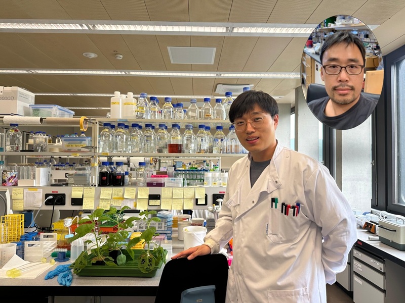
<Dr. Chaeyeon Lee, Professor Won Do Heo from Department of Biological Sciences>
A KAIST research team led by Professor Won Do Heo (Department of Biological Sciences) has developed an optogenetic platform, RELISR (REversible LIght-induced Store and Release), that enables precise spatiotemporal control over the storage and release of proteins and mRNAs in living cells and animals.
Traditional optogenetic condensate systems have been limited by their reliance on non-specific multivalent interactions, which can lead to unintended sequestration or release of endogenous molecules. RELISR overcomes these limitations by employing highly specific protein–protein (nanobody–antigen) and protein–RNA (MCP–MS2) interactions, enabling the selective and reversible compartmentalization of target proteins or mRNAs within engineered, membrane-less condensates.
In the dark, RELISR stably sequesters target molecules within condensates, physically isolating them from the cellular environment. Upon blue light stimulation, the condensates rapidly dissolve, releasing the stored proteins or mRNAs, which immediately regain their cellular functions or translational competency. This allows for reversible and rapid modulation of molecular activities in response to optical cues.
< Figure 1. Overview of the Artificial Condensate System (RELISR). The artificial condensate system, RELISR, includes "Protein-RELISR" for storing proteins and "mRNA-RELISR" for storing mRNA. These artificial condensates can be disassembled by blue light irradiation and reassembled in a dark state>
The research team demonstrated that RELISR enables temporal and spatial regulation of protein activity and mRNA translation in various cell types, including cultured neurons and mouse liver tissue. Comparative studies showed that RELISR provides more robust and reversible control of translation than previous systems based on spatial translocation.
While previous optogenetic systems such as LARIAT (Lee et al., Nature Methods, 2014) and mRNA-LARIAT (Kim et al., Nat. Cell Biol., 2019) enabled the selective sequestration of proteins or mRNAs into membrane-less condensates in response to light, they were primarily limited to the trapping phase. The RELISR platform introduced in this study establishes a new paradigm by enabling both the targeted storage of proteins and mRNAs and their rapid, light-triggered release. This approach allows researchers to not only confine molecular function on demand, but also to restore activity with precise temporal control.
< Figure 2. Cell shape change using the artificial condensate system (RELISR). A target protein, Vav2, which contributes to cell shape, was stored within the artificial condensate and then released after light irradiation. This release activated the target protein Vav2, causing a change in cell shape. It was confirmed that the storage, release, and activation of various proteins were effectively achieved>
Professor Heo stated, “RELISR is a versatile optogenetic tool that enables the precise control of protein and mRNA function at defined times and locations in living systems. We anticipate this platform will be broadly applicable for studies of cell signaling, neural circuits, and therapeutic development. Furthermore, the combination of RELISR with genome editing or tissue-targeted delivery could further expand its utility for molecular medicine.”
< Figure 3. Expression of a target mRNA using the artificial condensate system (RELISR) in mice. The genetic material for the artificial condensate system, RELISR, was injected into a living mouse. Using this system, a target mRNA was stored within the mouse's liver. Upon light irradiation, the mRNA was released, which induced the translation of a luminescent protein>
This research was conducted by first author Dr. Chaeyeon Lee, under the supervision of Professor Heo, with contributions from Dr. Daseuli Yu (co-corresponding author) and Professor YongKeun Park (co-corresponding author, Department of Physics), whose group performed quantitative imaging analyses of biophysical changes induced by RELISR in cells.
The findings were published in Nature Communications (July 7, 2025; DOI: 10.1038/s41467-025-61322-y). This work was supported by the Samsung Future Technology Foundation and the National Research Foundation of Korea.
2025.07.23 View 4122
KAIST Team Develops Optogenetic Platform for Spatiotemporal Control of Protein and mRNA Storage and Release
<Dr. Chaeyeon Lee, Professor Won Do Heo from Department of Biological Sciences>
A KAIST research team led by Professor Won Do Heo (Department of Biological Sciences) has developed an optogenetic platform, RELISR (REversible LIght-induced Store and Release), that enables precise spatiotemporal control over the storage and release of proteins and mRNAs in living cells and animals.
Traditional optogenetic condensate systems have been limited by their reliance on non-specific multivalent interactions, which can lead to unintended sequestration or release of endogenous molecules. RELISR overcomes these limitations by employing highly specific protein–protein (nanobody–antigen) and protein–RNA (MCP–MS2) interactions, enabling the selective and reversible compartmentalization of target proteins or mRNAs within engineered, membrane-less condensates.
In the dark, RELISR stably sequesters target molecules within condensates, physically isolating them from the cellular environment. Upon blue light stimulation, the condensates rapidly dissolve, releasing the stored proteins or mRNAs, which immediately regain their cellular functions or translational competency. This allows for reversible and rapid modulation of molecular activities in response to optical cues.
< Figure 1. Overview of the Artificial Condensate System (RELISR). The artificial condensate system, RELISR, includes "Protein-RELISR" for storing proteins and "mRNA-RELISR" for storing mRNA. These artificial condensates can be disassembled by blue light irradiation and reassembled in a dark state>
The research team demonstrated that RELISR enables temporal and spatial regulation of protein activity and mRNA translation in various cell types, including cultured neurons and mouse liver tissue. Comparative studies showed that RELISR provides more robust and reversible control of translation than previous systems based on spatial translocation.
While previous optogenetic systems such as LARIAT (Lee et al., Nature Methods, 2014) and mRNA-LARIAT (Kim et al., Nat. Cell Biol., 2019) enabled the selective sequestration of proteins or mRNAs into membrane-less condensates in response to light, they were primarily limited to the trapping phase. The RELISR platform introduced in this study establishes a new paradigm by enabling both the targeted storage of proteins and mRNAs and their rapid, light-triggered release. This approach allows researchers to not only confine molecular function on demand, but also to restore activity with precise temporal control.
< Figure 2. Cell shape change using the artificial condensate system (RELISR). A target protein, Vav2, which contributes to cell shape, was stored within the artificial condensate and then released after light irradiation. This release activated the target protein Vav2, causing a change in cell shape. It was confirmed that the storage, release, and activation of various proteins were effectively achieved>
Professor Heo stated, “RELISR is a versatile optogenetic tool that enables the precise control of protein and mRNA function at defined times and locations in living systems. We anticipate this platform will be broadly applicable for studies of cell signaling, neural circuits, and therapeutic development. Furthermore, the combination of RELISR with genome editing or tissue-targeted delivery could further expand its utility for molecular medicine.”
< Figure 3. Expression of a target mRNA using the artificial condensate system (RELISR) in mice. The genetic material for the artificial condensate system, RELISR, was injected into a living mouse. Using this system, a target mRNA was stored within the mouse's liver. Upon light irradiation, the mRNA was released, which induced the translation of a luminescent protein>
This research was conducted by first author Dr. Chaeyeon Lee, under the supervision of Professor Heo, with contributions from Dr. Daseuli Yu (co-corresponding author) and Professor YongKeun Park (co-corresponding author, Department of Physics), whose group performed quantitative imaging analyses of biophysical changes induced by RELISR in cells.
The findings were published in Nature Communications (July 7, 2025; DOI: 10.1038/s41467-025-61322-y). This work was supported by the Samsung Future Technology Foundation and the National Research Foundation of Korea.
2025.07.23 View 4122 -
 Why Do Plants Attack Themselves? The Secret of Genetic Conflict Revealed
<Professor Ji-Joon Song of the KAIST Department of Biological Sciences>
Plants, with their unique immune systems, sometimes launch 'autoimmune responses' by mistakenly identifying their own protein structures as pathogens. In particular, 'hybrid necrosis,' a phenomenon where descendant plants fail to grow healthily and perish after cross-breeding different varieties, has long been a difficult challenge for botanists and agricultural researchers. In response, an international research team has successfully elucidated the mechanism inducing plant autoimmune responses and proposed a novel strategy for cultivar improvement that can predict and avoid these reactions.
Professor Ji-Joon Song's research team at KAIST, in collaboration with teams from the National University of Singapore (NUS) and the University of Oxford, announced on the 21st of July that they have elucidated the structure and function of the 'DM3' protein complex, which triggers plant autoimmune responses, using cryo-electron microscopy (Cryo-EM) technology.
This research is drawing attention because it identifies defects in protein structure as the cause of hybrid necrosis, which occurs due to an abnormal reaction of immune receptors during cross-breeding between plant hybrids.
This protein (DM3) is originally an enzyme involved in the plant's immune response, but problems arise when the structure of the DM3 protein is damaged in a specific protein combination called 'DANGEROUS MIX (DM)'.
Notably, one variant of DM3, the 'DM3Col-0' variant, forms a stable complex with six proteins and is recognized as normal, thus not triggering an immune response. In contrast, another 'DM3Hh-0' variant has improper binding between its six proteins, causing the plant to recognize it as an 'abnormal state' and trigger an immune alarm, leading to autoimmunity.
The research team visualized this structure using atomic-resolution cryo-electron microscopy (Cryo-EM) and revealed that the immune-inducing ability is not due to the enzymatic function of the DM3 protein, but rather to 'differences in protein binding affinity.'
<Figure 1. Mechanism of Plant Autoimmunity Triggered by the Collapse of the DM3 Protein Complex>
This demonstrates that plants can initiate an immune response by recognizing not only 'external pathogens' but also 'internal protein structures' when they undergo abnormal changes, treating them as if they were pathogens.
The study shows how sensitively the plant immune system changes and triggers autoimmune responses when genes are mixed and protein structures change during the cross-breeding of different plant varieties. It significantly advanced the understanding of genetic incompatibility that can occur during natural cross-breeding and cultivar improvement processes.
Dr. Gijeong Kim, the co-first author, stated, "Through international research collaboration, we presented a new perspective on understanding the plant immune system by leveraging the autoimmune phenomenon, completing a high-quality study that encompasses structural biochemistry, genetics, and cell biological experiments."
Professor Ji-Joon Song of the KAIST Department of Biological Sciences, who led the research, said, "The fact that the immune system can detect not only external pathogens but also structural abnormalities in its own proteins will set a new standard for plant biotechnology and crop breeding strategies. Cryo-electron microscopy-based structural analysis will be an important tool for understanding the essence of gene interactions."
This research, with Professor Ji-Joon Song and Professor Eunyoung Chae of the University of Oxford as co-corresponding authors, Dr. Gijeong Kim (currently a postdoctoral researcher at the University of Zurich) and Dr. Wei-Lin Wan of the National University of Singapore as co-first authors, and Ph.D candidate Nayun Kim, as the second author, was published on July 17th in Molecular Cell, a sister journal of the international academic journal Cell.
This research was supported by the KAIST Grand Challenge 30 project.
Article Title: Structural determinants of DANGEROUS MIX 3, an alpha/beta hydrolase that triggers NLR-mediated genetic incompatibility in plants DOI: https://doi.org/10.1016/j.molcel.2025.06.021
2025.07.21 View 4191
Why Do Plants Attack Themselves? The Secret of Genetic Conflict Revealed
<Professor Ji-Joon Song of the KAIST Department of Biological Sciences>
Plants, with their unique immune systems, sometimes launch 'autoimmune responses' by mistakenly identifying their own protein structures as pathogens. In particular, 'hybrid necrosis,' a phenomenon where descendant plants fail to grow healthily and perish after cross-breeding different varieties, has long been a difficult challenge for botanists and agricultural researchers. In response, an international research team has successfully elucidated the mechanism inducing plant autoimmune responses and proposed a novel strategy for cultivar improvement that can predict and avoid these reactions.
Professor Ji-Joon Song's research team at KAIST, in collaboration with teams from the National University of Singapore (NUS) and the University of Oxford, announced on the 21st of July that they have elucidated the structure and function of the 'DM3' protein complex, which triggers plant autoimmune responses, using cryo-electron microscopy (Cryo-EM) technology.
This research is drawing attention because it identifies defects in protein structure as the cause of hybrid necrosis, which occurs due to an abnormal reaction of immune receptors during cross-breeding between plant hybrids.
This protein (DM3) is originally an enzyme involved in the plant's immune response, but problems arise when the structure of the DM3 protein is damaged in a specific protein combination called 'DANGEROUS MIX (DM)'.
Notably, one variant of DM3, the 'DM3Col-0' variant, forms a stable complex with six proteins and is recognized as normal, thus not triggering an immune response. In contrast, another 'DM3Hh-0' variant has improper binding between its six proteins, causing the plant to recognize it as an 'abnormal state' and trigger an immune alarm, leading to autoimmunity.
The research team visualized this structure using atomic-resolution cryo-electron microscopy (Cryo-EM) and revealed that the immune-inducing ability is not due to the enzymatic function of the DM3 protein, but rather to 'differences in protein binding affinity.'
<Figure 1. Mechanism of Plant Autoimmunity Triggered by the Collapse of the DM3 Protein Complex>
This demonstrates that plants can initiate an immune response by recognizing not only 'external pathogens' but also 'internal protein structures' when they undergo abnormal changes, treating them as if they were pathogens.
The study shows how sensitively the plant immune system changes and triggers autoimmune responses when genes are mixed and protein structures change during the cross-breeding of different plant varieties. It significantly advanced the understanding of genetic incompatibility that can occur during natural cross-breeding and cultivar improvement processes.
Dr. Gijeong Kim, the co-first author, stated, "Through international research collaboration, we presented a new perspective on understanding the plant immune system by leveraging the autoimmune phenomenon, completing a high-quality study that encompasses structural biochemistry, genetics, and cell biological experiments."
Professor Ji-Joon Song of the KAIST Department of Biological Sciences, who led the research, said, "The fact that the immune system can detect not only external pathogens but also structural abnormalities in its own proteins will set a new standard for plant biotechnology and crop breeding strategies. Cryo-electron microscopy-based structural analysis will be an important tool for understanding the essence of gene interactions."
This research, with Professor Ji-Joon Song and Professor Eunyoung Chae of the University of Oxford as co-corresponding authors, Dr. Gijeong Kim (currently a postdoctoral researcher at the University of Zurich) and Dr. Wei-Lin Wan of the National University of Singapore as co-first authors, and Ph.D candidate Nayun Kim, as the second author, was published on July 17th in Molecular Cell, a sister journal of the international academic journal Cell.
This research was supported by the KAIST Grand Challenge 30 project.
Article Title: Structural determinants of DANGEROUS MIX 3, an alpha/beta hydrolase that triggers NLR-mediated genetic incompatibility in plants DOI: https://doi.org/10.1016/j.molcel.2025.06.021
2025.07.21 View 4191 -
 KAIST Discovers Protein Switch that Turns Anti-Viral Immune Response On and Off
Even after the COVID-19 pandemic, various new infectious diseases continue to emerge, posing ongoing viral threats that demand robust and sustained immune defenses. However, excessive immune reactions can also harm body tissues, causing significant health issues. KAIST and an international research team have discovered a critical protein that acts as a 'switch' regulating immune responses to viruses. This breakthrough is expected to lay the groundwork for future infectious disease responses and autoimmune disease treatment strategies.
KAIST (President Kwang-Hyung Lee) announced on May 14 that a joint research team led by Professor Yoosik Kim from the Department of Chemical and Biomolecular Engineering at KAIST and Professor Seunghee Cha from University of Florida has discovered the mechanism by which double-stranded RNA derived from mitochondria amplifies immune responses. They identified the protein SLIRP as an 'immune switch' that regulates this process, playing a crucial role in both viral infections and autoimmune diseases.
< (From left) Master's candidate Yewon Yang, Professor Yoosik Kim and Ph.D. candidate Doyeong Ku of the Department of Chemical and Biomolecular Engineering >
Autoimmune diseases arise when the immune system fails to differentiate between external pathogens and the body's own molecules, leading to self-directed attacks. Despite extensive research, the precise causes of excessive inflammatory conditions like Sjögren’s syndrome and systemic lupus erythematosus remain unclear, and effective treatments are still limited.
To uncover the molecular mechanisms driving immune hyperactivation and to identify potential regulatory factors, the research team led by Professor Yoosik Kim focused on mitochondrial double-stranded RNA (mt-dsRNA), a genetic immunogenic material produced within cellular organelles. Since mt-dsRNA structurally resembles viral RNA, it can mistakenly trigger immune responses even in the absence of an actual viral infection.
The team discovered that SLIRP, a key regulator of mt-dsRNA, amplifies immune responses by stabilizing the RNA. They confirmed that SLIRP expression increases in experimental models simulating the tissues of autoimmune disease patients and viral infections. Conversely, suppressing SLIRP significantly reduced the immune response, underscoring its role as a critical factor in immune amplification.
This study also demonstrated the dual function of SLIRP in different contexts. In cells infected with human beta coronavirus OC43 and encephalomyocarditis virus (EMCV), SLIRP suppression led to reduced antiviral responses and increased viral replication. Meanwhile, in the blood and salivary gland cells of Sjögren’s syndrome patients, where both SLIRP and mt-dsRNA levels were elevated, suppressing SLIRP alleviated the abnormal immune response.
These findings highlight SLIRP as a key molecular switch that regulates immune responses in both infections and autoimmune diseases.
< Figure 1. Schematic diagram of antiviral signal amplification by SLIRP: SLIRP-based mt-dsRNA induction, cytoplasmic accumulation, and strong interferon response induction by positive feedback of immune response activation. Confirmation of the immune regulatory function of SLIRP in defense against autoimmune diseases Sjögren's syndrome, coronavirus, and encephalomyocarditis virus infection. >
Professor Yoosik Kim remarked, "Through this study, we have identified SLIRP as a crucial protein that drives immune amplification via mt-dsRNAs. Given its dual role in autoimmune diseases and viral infections, SLIRP presents a promising target for immune regulation therapies across various inflammatory disease contexts."
The study, with Ph.D. student Do-Young Ku (first author) and M.S. student Ye-Won Yang (second author) from the Department of Chemical and Biomolecular Engineering at KAIST as primary contributors, was published online in the journal Cell Reports on April 19, 2025.
※ Paper title: SLIRP amplifies antiviral signaling via positive feedback regulation and contributes to autoimmune diseases※ Main authors: Do-Young Ku (KAIST, first author), Ye-Won Yang (KAIST, second author), Seunghee Cha (University of Florida, corresponding author), Yoosik Kim (KAIST, corresponding author)
This study was supported by the Ministry of Health and Welfare's Public Health Technology Research Program and the National Institutes of Health (NIH) through Research Project (R01) funding.
2025.05.14 View 8269
KAIST Discovers Protein Switch that Turns Anti-Viral Immune Response On and Off
Even after the COVID-19 pandemic, various new infectious diseases continue to emerge, posing ongoing viral threats that demand robust and sustained immune defenses. However, excessive immune reactions can also harm body tissues, causing significant health issues. KAIST and an international research team have discovered a critical protein that acts as a 'switch' regulating immune responses to viruses. This breakthrough is expected to lay the groundwork for future infectious disease responses and autoimmune disease treatment strategies.
KAIST (President Kwang-Hyung Lee) announced on May 14 that a joint research team led by Professor Yoosik Kim from the Department of Chemical and Biomolecular Engineering at KAIST and Professor Seunghee Cha from University of Florida has discovered the mechanism by which double-stranded RNA derived from mitochondria amplifies immune responses. They identified the protein SLIRP as an 'immune switch' that regulates this process, playing a crucial role in both viral infections and autoimmune diseases.
< (From left) Master's candidate Yewon Yang, Professor Yoosik Kim and Ph.D. candidate Doyeong Ku of the Department of Chemical and Biomolecular Engineering >
Autoimmune diseases arise when the immune system fails to differentiate between external pathogens and the body's own molecules, leading to self-directed attacks. Despite extensive research, the precise causes of excessive inflammatory conditions like Sjögren’s syndrome and systemic lupus erythematosus remain unclear, and effective treatments are still limited.
To uncover the molecular mechanisms driving immune hyperactivation and to identify potential regulatory factors, the research team led by Professor Yoosik Kim focused on mitochondrial double-stranded RNA (mt-dsRNA), a genetic immunogenic material produced within cellular organelles. Since mt-dsRNA structurally resembles viral RNA, it can mistakenly trigger immune responses even in the absence of an actual viral infection.
The team discovered that SLIRP, a key regulator of mt-dsRNA, amplifies immune responses by stabilizing the RNA. They confirmed that SLIRP expression increases in experimental models simulating the tissues of autoimmune disease patients and viral infections. Conversely, suppressing SLIRP significantly reduced the immune response, underscoring its role as a critical factor in immune amplification.
This study also demonstrated the dual function of SLIRP in different contexts. In cells infected with human beta coronavirus OC43 and encephalomyocarditis virus (EMCV), SLIRP suppression led to reduced antiviral responses and increased viral replication. Meanwhile, in the blood and salivary gland cells of Sjögren’s syndrome patients, where both SLIRP and mt-dsRNA levels were elevated, suppressing SLIRP alleviated the abnormal immune response.
These findings highlight SLIRP as a key molecular switch that regulates immune responses in both infections and autoimmune diseases.
< Figure 1. Schematic diagram of antiviral signal amplification by SLIRP: SLIRP-based mt-dsRNA induction, cytoplasmic accumulation, and strong interferon response induction by positive feedback of immune response activation. Confirmation of the immune regulatory function of SLIRP in defense against autoimmune diseases Sjögren's syndrome, coronavirus, and encephalomyocarditis virus infection. >
Professor Yoosik Kim remarked, "Through this study, we have identified SLIRP as a crucial protein that drives immune amplification via mt-dsRNAs. Given its dual role in autoimmune diseases and viral infections, SLIRP presents a promising target for immune regulation therapies across various inflammatory disease contexts."
The study, with Ph.D. student Do-Young Ku (first author) and M.S. student Ye-Won Yang (second author) from the Department of Chemical and Biomolecular Engineering at KAIST as primary contributors, was published online in the journal Cell Reports on April 19, 2025.
※ Paper title: SLIRP amplifies antiviral signaling via positive feedback regulation and contributes to autoimmune diseases※ Main authors: Do-Young Ku (KAIST, first author), Ye-Won Yang (KAIST, second author), Seunghee Cha (University of Florida, corresponding author), Yoosik Kim (KAIST, corresponding author)
This study was supported by the Ministry of Health and Welfare's Public Health Technology Research Program and the National Institutes of Health (NIH) through Research Project (R01) funding.
2025.05.14 View 8269 -
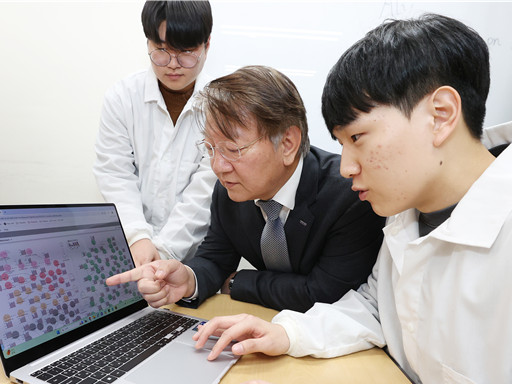 KAIST Accelerates Synthetic Microbe Design by Discovering Novel Enzymes Using AI
< (From left) Professor Sang Yup Lee of the Department of Chemical and Biomolecular Engineering (top), Hongkeun Ji, PhD candidate of the Department of Chemical and Biomolecular Engineering (top), Ha Rim Kim, PhD candidate of the Department of Chemical and Biomolecular Engineering, and Dr. Gi Bae Kim of the BioProcess Engineering Research Center >
Enzymes are proteins that catalyze biochemical reactions within cells and play a pivotal role in metabolic processes. Accordingly, identifying the functions of novel enzymes is a critical task in the construction of microbial cell factories.
A KAIST research team has leveraged artificial intelligence (AI) to design novel enzymes that do not exist in nature, significantly accelerating microbial cell factory development and boosting the potential for next-generation biotechnological applications such as drug development and biofuel production.
KAIST (represented by President Kwang-Hyung Lee) announced on the 21st of April that Distinguished Professor Sang Yup Lee and his team from the Department of Chemical and Biomolecular Engineering have published a review titled “Enzyme Functional Classification Using Artificial Intelligence,” which outlines the advancement of AI-based enzyme function prediction technologies and analyzes how AI has contributed to the discovery and design of new enzymes.
Professor Lee’s team systematically reviewed the development of enzyme function prediction technologies utilizing machine learning and deep learning, offering a comprehensive analysis.
From sequence similarity-based prediction methods to the integration of convolutional neural networks (CNNs), recurrent neural networks (RNNs), graph neural networks (GNNs), and transformer-based large language models, the paper covers a broad range of AI applications. It analyzes how these technologies extract meaningful information from protein sequences and enhance prediction accuracy.
In particular, enzyme function prediction using deep learning goes beyond simple sequence similarity analysis. By automatically extracting structural and evolutionary features embedded in amino acid sequences, deep learning enables more precise predictions of catalytic functions.
This highlights the unique advantages of AI models compared to traditional bioinformatics approaches.
Moreover, the review suggests that the advancement of generative AI will move future research beyond predicting existing functions to generating entirely new enzymes with functions not found in nature. This shift is expected to profoundly impact the trajectory of biotechnology and synthetic biology.
< Figure 1. Extraction of enzyme characteristics and function prediction using various deep learning structures >
Ha Rim Kim, a Ph.D. candidate and co-first author from the Department of Chemical and Biomolecular Engineering, stated, “AI-based enzyme function prediction and enzyme design are highly important across various fields including metabolic engineering, synthetic biology, and healthcare.”
Distinguished Professor Sang Yup Lee added, “AI-powered enzyme function prediction shows the potential to solve diverse biological problems and will significantly contribute to accelerating research across the entire field.”
The review was published on March 28 in Trends in Biotechnology, a leading biotechnology journal issued by Cell Press.
※ Title: Enzyme Functional Classification Using Artificial Intelligence
※DOI: https://doi.org/10.1016/j.tibtech.2025.03.003
※ Author Information: Ha Rim Kim (KAIST, Co-first author), Hongkeun Ji (KAIST, Co-first author), Gi Bae Kim (KAIST, Third author), Sang Yup Lee (KAIST, Corresponding author)
This research was supported by the Ministry of Science and ICT under the project Development of Core Technologies for Advanced Synthetic Biology to Lead the Bio-Manufacturing Industry (aimed at replacing petroleum-based chemicals), and also by joint support from the Ministry of Science and ICT and the Ministry of Health and Welfare for the project Development of Novel Antibiotic Structures Using Deep Learning-Based Synthetic Biology.
2025.04.07 View 10558
KAIST Accelerates Synthetic Microbe Design by Discovering Novel Enzymes Using AI
< (From left) Professor Sang Yup Lee of the Department of Chemical and Biomolecular Engineering (top), Hongkeun Ji, PhD candidate of the Department of Chemical and Biomolecular Engineering (top), Ha Rim Kim, PhD candidate of the Department of Chemical and Biomolecular Engineering, and Dr. Gi Bae Kim of the BioProcess Engineering Research Center >
Enzymes are proteins that catalyze biochemical reactions within cells and play a pivotal role in metabolic processes. Accordingly, identifying the functions of novel enzymes is a critical task in the construction of microbial cell factories.
A KAIST research team has leveraged artificial intelligence (AI) to design novel enzymes that do not exist in nature, significantly accelerating microbial cell factory development and boosting the potential for next-generation biotechnological applications such as drug development and biofuel production.
KAIST (represented by President Kwang-Hyung Lee) announced on the 21st of April that Distinguished Professor Sang Yup Lee and his team from the Department of Chemical and Biomolecular Engineering have published a review titled “Enzyme Functional Classification Using Artificial Intelligence,” which outlines the advancement of AI-based enzyme function prediction technologies and analyzes how AI has contributed to the discovery and design of new enzymes.
Professor Lee’s team systematically reviewed the development of enzyme function prediction technologies utilizing machine learning and deep learning, offering a comprehensive analysis.
From sequence similarity-based prediction methods to the integration of convolutional neural networks (CNNs), recurrent neural networks (RNNs), graph neural networks (GNNs), and transformer-based large language models, the paper covers a broad range of AI applications. It analyzes how these technologies extract meaningful information from protein sequences and enhance prediction accuracy.
In particular, enzyme function prediction using deep learning goes beyond simple sequence similarity analysis. By automatically extracting structural and evolutionary features embedded in amino acid sequences, deep learning enables more precise predictions of catalytic functions.
This highlights the unique advantages of AI models compared to traditional bioinformatics approaches.
Moreover, the review suggests that the advancement of generative AI will move future research beyond predicting existing functions to generating entirely new enzymes with functions not found in nature. This shift is expected to profoundly impact the trajectory of biotechnology and synthetic biology.
< Figure 1. Extraction of enzyme characteristics and function prediction using various deep learning structures >
Ha Rim Kim, a Ph.D. candidate and co-first author from the Department of Chemical and Biomolecular Engineering, stated, “AI-based enzyme function prediction and enzyme design are highly important across various fields including metabolic engineering, synthetic biology, and healthcare.”
Distinguished Professor Sang Yup Lee added, “AI-powered enzyme function prediction shows the potential to solve diverse biological problems and will significantly contribute to accelerating research across the entire field.”
The review was published on March 28 in Trends in Biotechnology, a leading biotechnology journal issued by Cell Press.
※ Title: Enzyme Functional Classification Using Artificial Intelligence
※DOI: https://doi.org/10.1016/j.tibtech.2025.03.003
※ Author Information: Ha Rim Kim (KAIST, Co-first author), Hongkeun Ji (KAIST, Co-first author), Gi Bae Kim (KAIST, Third author), Sang Yup Lee (KAIST, Corresponding author)
This research was supported by the Ministry of Science and ICT under the project Development of Core Technologies for Advanced Synthetic Biology to Lead the Bio-Manufacturing Industry (aimed at replacing petroleum-based chemicals), and also by joint support from the Ministry of Science and ICT and the Ministry of Health and Welfare for the project Development of Novel Antibiotic Structures Using Deep Learning-Based Synthetic Biology.
2025.04.07 View 10558 -
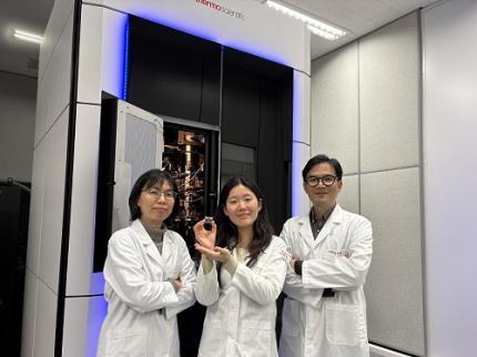 KAIST Captures Protein Reaction in Just Six Milliseconds
Understanding biomolecular processes - such as protein-protein interactions and enzyme-substrate reactions that occur on the microseconds to millisecond time scale is essential for comprehending life processes and advancing drug development. KAIST researchers have developed a method for freezing and analyzing biochemical reaction dynamics within a span of just a few milliseconds, marking a significant step forward in better understanding complex biological reactions.
< Photo. (From left) Professor Jin Young Kang and Haerang Hwang of the Integrated Master's and Doctoral Program of the Department of Chemistry, along with Professor Wonhee Lee of the Department of Physics >
KAIST (represented by President Kwang Hyung Lee) announced on the 24th of March that a joint research team led by Professor Jin Young Kang from the Department of Chemistry and Professor Wonhee Lee from the Department of Physics has developed a parylene-based thin-film microfluidic mixing-and-spraying device for ultra-fast biochemical reaction studies.
*Parylene: A key material for microfluidic devices used to observe protein dynamics at ultra-high speeds. It can be fabricated into a few micrometer-thick films, which can be used in making a spray nozzle for microfluidic devices.
This research overcomes the limitations of the existing time-resolved cryo-electron microscopy (TRCEM) method by reducing sample consumption to one-third of the conventional amount while improving the minimum time resolution—down to just six milliseconds (6 ms).
TRCEM is a technique that rapidly freezes protein complexes during intermediate reaction stages under cryogenic conditions, which allows researchers to analyze their structures. This approach has gained significant attention recently for its ability to capture transient biochemical events.
< Figure 1. Time-resolved cryo-EM (TRCEM) technique using microfluidic channels. In order to capture the intermediate structure of biomolecules during a biochemical reaction over time, biomolecules and reaction substrates are mixed in a microfluidic channel, and then sprayed on a grid after a certain reaction time and frozen in liquid ethane to prepare a cryo-EM sample. This can then be analyzed by cryo-EM to observe the structural changes of proteins over time. >
Transient intermediate structures of protein complexes could not be captured by traditional cryo-electron microscopy due to their extremely short lifespans. Although several TRCEM techniques have been developed to address this issue, previous methods were hindered by large sample consumption and limited time resolution. To overcome these challenges, the KAIST team developed a new mixing-and-spraying device using ultra-thin parylene films. The integrated design of the device further enhanced the precision and reproducibility of experiments.
< Figure 2. TRCEM grid fabrication setup using a parylene-based thin-film microfluidic device and actual appearance of the device. You can see that a thin-film parylene channel is inserted into the injection nozzle. The integration of the reaction channel and the injection nozzle allowed the residence time in the device to be reduced to at least 0.5 ms. >
“This research makes TRCEM more practical and paves the way for diverse applications of the parylene thin-film device in structural biology, drug development, enzyme reaction studies, and biosensor research.” Professor Jin Young Kang explained, emphasizing the significance of the study.
Professor Wonhee Lee added, “The team aims to continue this research, focusing on improvement of the technique to achieve higher time resolution with minimal sample consumption.”
< Figure 3. Comparison of the spraying patterns of the parylene mixing-jet device and the conventional mixing-jet device and the filament length in the resulting RecA-ssDNA filament formation reaction. It was shown that the thin film spray nozzle structure affects the uniformity and accuracy of the final reaction time. >
The research findings, with Haerang Hwang (a graduate student in the integrated master's and Ph.D. program in the Department of Chemistry) as the first author, were published online on January 28, 2025, in the international journal Advanced Functional Materials. (Paper Title: “Integrated Parylene-Based Thin-Film Microfluidic Device for Time-Resolved Cryo-Electron Microscopy”, DOI: doi.org/10.1002/adfm.202418224)
This research was supported by the National Research Foundation of Korea (NRF), the Samsung Future Technology Development Program, and the CELINE consortium.
2025.03.24 View 9788
KAIST Captures Protein Reaction in Just Six Milliseconds
Understanding biomolecular processes - such as protein-protein interactions and enzyme-substrate reactions that occur on the microseconds to millisecond time scale is essential for comprehending life processes and advancing drug development. KAIST researchers have developed a method for freezing and analyzing biochemical reaction dynamics within a span of just a few milliseconds, marking a significant step forward in better understanding complex biological reactions.
< Photo. (From left) Professor Jin Young Kang and Haerang Hwang of the Integrated Master's and Doctoral Program of the Department of Chemistry, along with Professor Wonhee Lee of the Department of Physics >
KAIST (represented by President Kwang Hyung Lee) announced on the 24th of March that a joint research team led by Professor Jin Young Kang from the Department of Chemistry and Professor Wonhee Lee from the Department of Physics has developed a parylene-based thin-film microfluidic mixing-and-spraying device for ultra-fast biochemical reaction studies.
*Parylene: A key material for microfluidic devices used to observe protein dynamics at ultra-high speeds. It can be fabricated into a few micrometer-thick films, which can be used in making a spray nozzle for microfluidic devices.
This research overcomes the limitations of the existing time-resolved cryo-electron microscopy (TRCEM) method by reducing sample consumption to one-third of the conventional amount while improving the minimum time resolution—down to just six milliseconds (6 ms).
TRCEM is a technique that rapidly freezes protein complexes during intermediate reaction stages under cryogenic conditions, which allows researchers to analyze their structures. This approach has gained significant attention recently for its ability to capture transient biochemical events.
< Figure 1. Time-resolved cryo-EM (TRCEM) technique using microfluidic channels. In order to capture the intermediate structure of biomolecules during a biochemical reaction over time, biomolecules and reaction substrates are mixed in a microfluidic channel, and then sprayed on a grid after a certain reaction time and frozen in liquid ethane to prepare a cryo-EM sample. This can then be analyzed by cryo-EM to observe the structural changes of proteins over time. >
Transient intermediate structures of protein complexes could not be captured by traditional cryo-electron microscopy due to their extremely short lifespans. Although several TRCEM techniques have been developed to address this issue, previous methods were hindered by large sample consumption and limited time resolution. To overcome these challenges, the KAIST team developed a new mixing-and-spraying device using ultra-thin parylene films. The integrated design of the device further enhanced the precision and reproducibility of experiments.
< Figure 2. TRCEM grid fabrication setup using a parylene-based thin-film microfluidic device and actual appearance of the device. You can see that a thin-film parylene channel is inserted into the injection nozzle. The integration of the reaction channel and the injection nozzle allowed the residence time in the device to be reduced to at least 0.5 ms. >
“This research makes TRCEM more practical and paves the way for diverse applications of the parylene thin-film device in structural biology, drug development, enzyme reaction studies, and biosensor research.” Professor Jin Young Kang explained, emphasizing the significance of the study.
Professor Wonhee Lee added, “The team aims to continue this research, focusing on improvement of the technique to achieve higher time resolution with minimal sample consumption.”
< Figure 3. Comparison of the spraying patterns of the parylene mixing-jet device and the conventional mixing-jet device and the filament length in the resulting RecA-ssDNA filament formation reaction. It was shown that the thin film spray nozzle structure affects the uniformity and accuracy of the final reaction time. >
The research findings, with Haerang Hwang (a graduate student in the integrated master's and Ph.D. program in the Department of Chemistry) as the first author, were published online on January 28, 2025, in the international journal Advanced Functional Materials. (Paper Title: “Integrated Parylene-Based Thin-Film Microfluidic Device for Time-Resolved Cryo-Electron Microscopy”, DOI: doi.org/10.1002/adfm.202418224)
This research was supported by the National Research Foundation of Korea (NRF), the Samsung Future Technology Development Program, and the CELINE consortium.
2025.03.24 View 9788 -
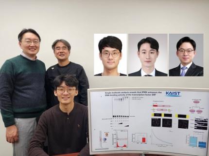 KAIST Uncovers the Principles of Gene Expression Regulation in Cancer and Cellular Functions
< (From left) Professor Seyun Kim, Professor Gwangrog Lee, Dr. Hyoungjoon Ahn, Dr. Jeongmin Yu, Professor Won-Ki Cho, and (below) PhD candidate Kwangmin Ryu of the Department of Biological Sciences>
A research team at KAIST has identified the core gene expression networks regulated by key proteins that fundamentally drive phenomena such as cancer development, metastasis, tissue differentiation from stem cells, and neural activation processes. This discovery lays the foundation for developing innovative therapeutic technologies.
On the 22nd of January, KAIST (represented by President Kwang Hyung Lee) announced that the joint research team led by Professors Seyun Kim, Gwangrog Lee, and Won-Ki Cho from the Department of Biological Sciences had uncovered essential mechanisms controlling gene expression in animal cells.
Inositol phosphate metabolites produced by inositol metabolism enzymes serve as vital secondary messengers in eukaryotic cell signaling systems and are broadly implicated in cancer, obesity, diabetes, and neurological disorders.
The research team demonstrated that the inositol polyphosphate multikinase (IPMK) enzyme, a key player in the inositol metabolism system, acts as a critical transcriptional activator within the core gene expression networks of animal cells. Notably, although IPMK was previously reported to play an important role in the transcription process governed by serum response factor (SRF), a representative transcription factor in animal cells, the precise mechanism of its action was unclear.
SRF is a transcription factor directly controlling the expression of at least 200–300 genes, regulating cell growth, proliferation, apoptosis, and motility, and is indispensable for organ development, such as in the heart.
The team discovered that IPMK binds directly to SRF, altering the three-dimensional structure of the SRF protein. This interaction facilitates the transcriptional activity of various genes through the SRF activated by IPMK, demonstrating that IPMK acts as a critical regulatory switch to enhance SRF's protein activity.
< Figure 1. The serum response factor (SRF) protein, a key transcription factor in animal cells, directly binds to inositol polyphosphate multikinase (IPMK) enzyme and undergoes structural change to acquire DNA binding ability, and precisely regulates growth and differentiation of animal cells through transcriptional activation. >
The team further verified that disruptions in the direct interaction between IPMK and SRF lead to the reduced functionality and activity of SRF, causing severe impairments in gene expression.
By highlighting the significance of the intrinsically disordered region (IDR) in SRF, the researchers underscored the biological importance of intrinsically disordered proteins (IDPs). Unlike most proteins that adopt distinct structures through folding, IDPs, including those with IDRs, do not exhibit specific structures but play crucial biological roles, attracting significant attention in the scientific community.
Professor Seyun Kim commented, "This study provides a vital mechanism proving that IPMK, a key enzyme in the inositol metabolism system, is a major transcriptional activator in the core gene expression network of animal cells. By understanding fundamental processes such as cancer development and metastasis, tissue differentiation from stem cells, and neural activation through SRF, we hope this discovery will lead to the broad application of innovative therapeutic technologies."
The findings were published on January 7th in the international journal Nucleic Acids Research (IF=16.7, top 1.8% in Biochemistry and Molecular Biology), under the title “Single-molecule analysis reveals that IPMK enhances the DNA-binding activity of the transcription factor SRF" (DOI: 10.1093/nar/gkae1281).
This research was supported by the National Research Foundation of Korea's Mid-career Research Program, Leading Research Center Program, and Global Research Laboratory Program, as well as by the Suh Kyungbae Science Foundation and the Samsung Future Technology Development Program.
2025.01.24 View 18638
KAIST Uncovers the Principles of Gene Expression Regulation in Cancer and Cellular Functions
< (From left) Professor Seyun Kim, Professor Gwangrog Lee, Dr. Hyoungjoon Ahn, Dr. Jeongmin Yu, Professor Won-Ki Cho, and (below) PhD candidate Kwangmin Ryu of the Department of Biological Sciences>
A research team at KAIST has identified the core gene expression networks regulated by key proteins that fundamentally drive phenomena such as cancer development, metastasis, tissue differentiation from stem cells, and neural activation processes. This discovery lays the foundation for developing innovative therapeutic technologies.
On the 22nd of January, KAIST (represented by President Kwang Hyung Lee) announced that the joint research team led by Professors Seyun Kim, Gwangrog Lee, and Won-Ki Cho from the Department of Biological Sciences had uncovered essential mechanisms controlling gene expression in animal cells.
Inositol phosphate metabolites produced by inositol metabolism enzymes serve as vital secondary messengers in eukaryotic cell signaling systems and are broadly implicated in cancer, obesity, diabetes, and neurological disorders.
The research team demonstrated that the inositol polyphosphate multikinase (IPMK) enzyme, a key player in the inositol metabolism system, acts as a critical transcriptional activator within the core gene expression networks of animal cells. Notably, although IPMK was previously reported to play an important role in the transcription process governed by serum response factor (SRF), a representative transcription factor in animal cells, the precise mechanism of its action was unclear.
SRF is a transcription factor directly controlling the expression of at least 200–300 genes, regulating cell growth, proliferation, apoptosis, and motility, and is indispensable for organ development, such as in the heart.
The team discovered that IPMK binds directly to SRF, altering the three-dimensional structure of the SRF protein. This interaction facilitates the transcriptional activity of various genes through the SRF activated by IPMK, demonstrating that IPMK acts as a critical regulatory switch to enhance SRF's protein activity.
< Figure 1. The serum response factor (SRF) protein, a key transcription factor in animal cells, directly binds to inositol polyphosphate multikinase (IPMK) enzyme and undergoes structural change to acquire DNA binding ability, and precisely regulates growth and differentiation of animal cells through transcriptional activation. >
The team further verified that disruptions in the direct interaction between IPMK and SRF lead to the reduced functionality and activity of SRF, causing severe impairments in gene expression.
By highlighting the significance of the intrinsically disordered region (IDR) in SRF, the researchers underscored the biological importance of intrinsically disordered proteins (IDPs). Unlike most proteins that adopt distinct structures through folding, IDPs, including those with IDRs, do not exhibit specific structures but play crucial biological roles, attracting significant attention in the scientific community.
Professor Seyun Kim commented, "This study provides a vital mechanism proving that IPMK, a key enzyme in the inositol metabolism system, is a major transcriptional activator in the core gene expression network of animal cells. By understanding fundamental processes such as cancer development and metastasis, tissue differentiation from stem cells, and neural activation through SRF, we hope this discovery will lead to the broad application of innovative therapeutic technologies."
The findings were published on January 7th in the international journal Nucleic Acids Research (IF=16.7, top 1.8% in Biochemistry and Molecular Biology), under the title “Single-molecule analysis reveals that IPMK enhances the DNA-binding activity of the transcription factor SRF" (DOI: 10.1093/nar/gkae1281).
This research was supported by the National Research Foundation of Korea's Mid-career Research Program, Leading Research Center Program, and Global Research Laboratory Program, as well as by the Suh Kyungbae Science Foundation and the Samsung Future Technology Development Program.
2025.01.24 View 18638