KAIST Uncovers the Mechanism Behind Overactive Immune Cells
<(From Right) Professor Eui-Cheol Shin, Ph.D candidate So-Young Kim, Professor Su-Hyung Park, Professor Hyuk Soo Eun, Dr. Hoyoung Lee>
“Why do immune cells that are supposed to eliminate viruses suddenly turn against our own body?”
There are instances where killer T cells—which are meant to precisely remove virus-infected cells—malfunction like overheated engines, attacking even healthy cells and damaging tissues. A KAIST research team has now identified the key mechanism that regulates this excessive activation of killer T cells, offering new insights into controlling immune overreactions and developing therapies for immune-related diseases.
KAIST (President Kwang Hyung Lee) announced on November 5 that a research team led by Professors Eui-Cheol Shin and Su-Hyung Park from the Graduate School of Medical Science and Engineering, in collaboration with Professor Hyuk Soo Eun from Chungnam National University College of Medicine, has uncovered the molecular basis of nonspecific activation in killer T cells and proposed a new therapeutic strategy to control it.
Killer T cells (CD8⁺ T cells) selectively eliminate infected cells to prevent viral spread. However, when excessively activated, they can attack uninfected cells, causing inflammation and tissue damage. Such overactive immune responses can lead to severe viral infections and autoimmune diseases.
In 2018, Professor Shin’s team was the first in the world to discover that killer T cells can be nonspecifically activated by cytokines and randomly attack host cells—a phenomenon they termed “bystander activation of T cells”. The current study builds on that discovery by revealing the molecular mechanism driving this abnormal process.
The team focused on a cytokine called interleukin-15 (IL-15). Experiments showed that IL-15 can abnormally excite killer T cells by a bystander activation mechanism, causing them to attack uninfected host cells. However, when there is a concurrent antigen-specific stimulation, IL-15-induced bystander activation is suppressed.
The researchers further identified that this suppression occurs through an intracellular signaling process. When the concentration of calcium ions (Ca²⁺) changes, a protein called calcineurin activates, which in turn triggers a regulatory protein known as NFAT, suppressing IL-15-induced bystander activation of killer T cells. In other words, the calcineurin–NFAT pathway activated by antigen stimulation acts as a brake on overactivation by a bystander mechanism.
The team also discovered that some immunosuppressants, which are known to block the calcineurin pathway, may not always suppress immune responses—in certain contexts, they can instead promote IL-15-induced bystander activation of killer T cells. This finding underscores that not all immunosuppressants work the same way and that treatments must be carefully tailored to each patient’s immune response.
Through gene expression analysis, the researchers identified a gene set that increase only in abnormally activated killer T cells induced by IL-15 as markers. They further confirmed that these same markers were elevated in bystander killer T cells from patients with acute hepatitis A, suggesting that the markers could be used for disease diagnosis.
<In a normal immune response, killer T cells are activated by antigen stimulation and selectively eliminate only virus-infected cells, thereby controlling viral replication and promoting the patient’s rapid recovery. However, when killer T cells are nonspecifically overactivated by interleukin-15, they may randomly attack normal cells as well, causing excessive tissue damage and leading to severe disease. Future research may identify diseases in which such nonspecific hyperimmune responses occur, making it possible to develop new drugs to control them>
This study provides crucial clues for understanding the pathogenesis of various immune-related diseases, including severe viral infections, chronic inflammatory disorders, autoimmune diseases, and organ transplant rejection. It also paves the way for developing novel immunoregulatory therapies targeting IL-15 signaling.
Professor Eui-Cheol Shin explained that, “this study shows that killer T cells are not merely defenders—they can transform into ‘nonspecific attackers’ depending on the inflammatory environment. By precisely regulating this abnormal activation, we may be able to develop new treatments for intractable immune diseases.”
This research was published in the journal Immunity on October 31, with Dr. Hoyoung Lee and Ph.D. candidate So-Young Kim as co–first authors.
Title: “TCR signaling via NFATc1 constrains IL-15-induced bystander activation of human memory CD8⁺ T cells”, DOI: doi.org/10.1016/j.immuni.2025.10.002
The study was supported by the National Research Foundation of Korea (NRF), the Korea Health Industry Development Institute (KHIDI), and the Institute for Basic Science (IBS).
Why Do Plants Attack Themselves? The Secret of Genetic Conflict Revealed
<Professor Ji-Joon Song of the KAIST Department of Biological Sciences>
Plants, with their unique immune systems, sometimes launch 'autoimmune responses' by mistakenly identifying their own protein structures as pathogens. In particular, 'hybrid necrosis,' a phenomenon where descendant plants fail to grow healthily and perish after cross-breeding different varieties, has long been a difficult challenge for botanists and agricultural researchers. In response, an international research team has successfully elucidated the mechanism inducing plant autoimmune responses and proposed a novel strategy for cultivar improvement that can predict and avoid these reactions.
Professor Ji-Joon Song's research team at KAIST, in collaboration with teams from the National University of Singapore (NUS) and the University of Oxford, announced on the 21st of July that they have elucidated the structure and function of the 'DM3' protein complex, which triggers plant autoimmune responses, using cryo-electron microscopy (Cryo-EM) technology.
This research is drawing attention because it identifies defects in protein structure as the cause of hybrid necrosis, which occurs due to an abnormal reaction of immune receptors during cross-breeding between plant hybrids.
This protein (DM3) is originally an enzyme involved in the plant's immune response, but problems arise when the structure of the DM3 protein is damaged in a specific protein combination called 'DANGEROUS MIX (DM)'.
Notably, one variant of DM3, the 'DM3Col-0' variant, forms a stable complex with six proteins and is recognized as normal, thus not triggering an immune response. In contrast, another 'DM3Hh-0' variant has improper binding between its six proteins, causing the plant to recognize it as an 'abnormal state' and trigger an immune alarm, leading to autoimmunity.
The research team visualized this structure using atomic-resolution cryo-electron microscopy (Cryo-EM) and revealed that the immune-inducing ability is not due to the enzymatic function of the DM3 protein, but rather to 'differences in protein binding affinity.'
<Figure 1. Mechanism of Plant Autoimmunity Triggered by the Collapse of the DM3 Protein Complex>
This demonstrates that plants can initiate an immune response by recognizing not only 'external pathogens' but also 'internal protein structures' when they undergo abnormal changes, treating them as if they were pathogens.
The study shows how sensitively the plant immune system changes and triggers autoimmune responses when genes are mixed and protein structures change during the cross-breeding of different plant varieties. It significantly advanced the understanding of genetic incompatibility that can occur during natural cross-breeding and cultivar improvement processes.
Dr. Gijeong Kim, the co-first author, stated, "Through international research collaboration, we presented a new perspective on understanding the plant immune system by leveraging the autoimmune phenomenon, completing a high-quality study that encompasses structural biochemistry, genetics, and cell biological experiments."
Professor Ji-Joon Song of the KAIST Department of Biological Sciences, who led the research, said, "The fact that the immune system can detect not only external pathogens but also structural abnormalities in its own proteins will set a new standard for plant biotechnology and crop breeding strategies. Cryo-electron microscopy-based structural analysis will be an important tool for understanding the essence of gene interactions."
This research, with Professor Ji-Joon Song and Professor Eunyoung Chae of the University of Oxford as co-corresponding authors, Dr. Gijeong Kim (currently a postdoctoral researcher at the University of Zurich) and Dr. Wei-Lin Wan of the National University of Singapore as co-first authors, and Ph.D candidate Nayun Kim, as the second author, was published on July 17th in Molecular Cell, a sister journal of the international academic journal Cell.
This research was supported by the KAIST Grand Challenge 30 project.
Article Title: Structural determinants of DANGEROUS MIX 3, an alpha/beta hydrolase that triggers NLR-mediated genetic incompatibility in plants DOI: https://doi.org/10.1016/j.molcel.2025.06.021
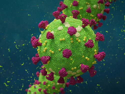
Biomarker Predicts Who Will Have Severe COVID-19
- Airway cell analyses showing an activated immune axis could pinpoint the COVID-19 patients who will most benefit from targeted therapies.-
KAIST researchers have identified key markers that could help pinpoint patients who are bound to get a severe reaction to COVID-19 infection. This would help doctors provide the right treatments at the right time, potentially saving lives. The findings were published in the journal Frontiers in Immunology on August 28.
People’s immune systems react differently to infection with SARS-CoV-2, the virus that causes COVID-19, ranging from mild to severe, life-threatening responses.
To understand the differences in responses, Professor Heung Kyu Lee and PhD candidate Jang Hyun Park from the Graduate School of Medical Science and Engineering at KAIST analysed ribonucleic acid (RNA) sequencing data extracted from individual airway cells of healthy controls and of mildly and severely ill patients with COVID-19. The data was available in a public database previously published by a group of Chinese researchers.
“Our analyses identified an association between immune cells called neutrophils and special cell receptors that bind to the steroid hormone glucocorticoid,” Professor Lee explained. “This finding could be used as a biomarker for predicting disease severity in patients and thus selecting a targeted therapy that can help treat them at an appropriate time,” he added.
Severe illness in COVID-19 is associated with an exaggerated immune response that leads to excessive airway-damaging inflammation. This condition, known as acute respiratory distress syndrome (ARDS), accounts for 70% of deaths in fatal COVID-19 infections.
Scientists already know that this excessive inflammation involves heightened neutrophil recruitment to the airways, but the detailed mechanisms of this reaction are still unclear.
Lee and Park’s analyses found that a group of immune cells called myeloid cells produced excess amounts of neutrophil-recruiting chemicals in severely ill patients, including a cytokine called tumour necrosis factor (TNF) and a chemokine called CXCL8.
Further RNA analyses of neutrophils in severely ill patients showed they were less able to recruit very important T cells needed for attacking the virus. At the same time, the neutrophils produced too many extracellular molecules that normally trap pathogens, but damage airway cells when produced in excess.
The researchers additionally found that the airway cells in severely ill patients were not expressing enough glucocorticoid receptors. This was correlated with increased CXCL8 expression and neutrophil recruitment.
Glucocorticoids, like the well-known drug dexamethasone, are anti-inflammatory agents that could play a role in treating COVID-19. However, using them in early or mild forms of the infection could suppress the necessary immune reactions to combat the virus. But if airway damage has already happened in more severe cases, glucocorticoid treatment would be ineffective.
Knowing who to give this treatment to and when is really important. COVID-19 patients showing reduced glucocorticoid receptor expression, increased CXCL8 expression, and excess neutrophil recruitment to the airways could benefit from treatment with glucocorticoids to prevent airway damage. Further research is needed, however, to confirm the relationship between glucocorticoids and neutrophil inflammation at the protein level.
“Our study could serve as a springboard towards more accurate and reliable COVID-19 treatments,” Professor Lee said.
This work was supported by the National Research Foundation of Korea, and Mobile Clinic Module Project funded by KAIST.
Figure. Low glucocorticoid receptor (GR) expression led to excessive inflammation and lung damage by neutrophils through enhancing the expression of CXCL8 and other cytokines.
Image credit: Professor Heung Kyu Lee, KAIST. Created with Biorender.com.
Image usage restrictions: News organizations may use or redistribute these figures and image, with proper attribution, as part of news coverage of this paper only.
-Publication:
Jang Hyun Park, and Heung Kyu Lee. (2020). Re-analysis of Single Cell Transcriptome Reveals That the NR3C1-CXCL8-Neutrophil Axis Determines the Severity of COVID-19. Frontiers in Immunology, Available online at https://doi.org/10.3389/fimmu.2020.02145
-Profile: Heung Kyu Lee
Associate Professor
heungkyu.lee@kaist.ac.kr
https://www.heungkyulee.kaist.ac.kr/
Laboratory of Host Defenses
Graduate School of Medical Science and Engineering (GSMSE)
The Center for Epidemic Preparedness at KAIST Institute
http://kaist.ac.kr
Korea Advanced Institute of Science and Technology (KAIST)
Daejeon, Republic of Korea
Profile: Jang Hyun Park
PhD Candidate
janghyun.park@kaist.ac.kr
GSMSE, KAIST