%54%72%65%61%74%6d%65%6e%74
-
 KAIST Proposes a New Dementia Treatment Strategy by Repositioning Molecules without Changing Their Chemical Composition
<(Back row, from left) Professor Mi Hee Lim, Professor Mingeun Kim, Student Jimin Lee, Student Chanju Na, (Upper Right) Dr. Chul-Ho Lee, Dr Kyoung-Shim Kim>
Conventional treatments of Alzheimer’s disease, one of the most common forms of dementia, have been largely focused on targeting individual pathological features. However, Alzheimer’s disease is a multifactorial disorder driven by multiple, tightly interconnected processes, rendering single-target therapeutic approaches inherently limited. Addressing this challenge, KAIST researchers propose a new strategy that enables the simultaneous regulation of multiple disease-inducing factors simply by rearranging the structural positions of drug candidate molecules without altering their chemical substituents.
KAIST (President Kwang Hyung Lee) announced on January 22 that a research team led by Professor Mi Hee Lim of the Department of Chemistry, in collaboration with Professor Mingeun Kim of Chonnam National University, Dr. Chul-Ho Lee of the Korea Research Institute of Bioscience and Biotechnology (KRIBB), and Dr. Kyoung-Shim Kim of the Laboratory Animal Resource Center, has elucidated at the molecular level how subtle differences in molecular arrangement, specifically positional isomerism, give rise to distinct modes of action against Alzheimer’s disease.
Using an Alzheimer’s disease mouse model (APP/PS1) harboring human dementia-associated genes, the research team demonstrated that these compounds also exert distinct therapeutic effects in vivo.
Alzheimer’s disease does not arise from a single cause. Rather, multiple pathological factors, including amyloid-b, metal ions, and reactive oxygen species, interact synergistically to exacerbate disease progression. In particular, metal ions bind to amyloid-b, modulating its aggregation and toxicity while promoting the generation of reactive oxygen species, which in turn accelerates neuronal damage. Effective control of Alzheimer’s disease therefore requires therapeutic strategies capable of simultaneously targeting multiple interrelated pathological processes.
< Alzheimer’s Disease – Chemical Approach Illustration (AI-generated image) >
The researchers focused on positional isomers, molecules composed of the same chemical elements but differing only in the positions at which those elements are connected. Remarkably, simple changes in molecular positioning resulted in pronounced differences in reactivity towards reactive oxygen species, as well as in interactions with amyloid-b and metal-bound amyloid-b.
To investigate these effects, the team compared the reactivities of three structurally similar molecules differing only in the positions of their functional groups. Their analyses revealed that even minimal structural rearrangements led to significant differences in antioxidant capacity and produced distinct modes of modulation of amyloid-b and metal-bound amyloid-b through different mechanisms, inducing peptide chemical modifications.
In other words, the study demonstrated that Alzheimer’s disease-related pathological factors can be regulated through mechanistically distinct pathways simply by altering molecular arrangement, without changing molecular composition.
Notably, a specific positional isomer capable of simultaneously modulating reactive oxygen species, amyloid-b, and metal-bound amyloid-b complexes also demonstrated therapeutic efficacy in an Alzheimer’s disease mouse model. In these experiments, the compound reduced oxidative stress in the hippocampus, the brain region critical for memory, and decreased amyloid plaque accumulation, resulting in significant improvements in memory deficits and cognitive impairment.
< In Vivo Efficacy Evaluation and Biological Outcomes According to Positional Isomers of Small-Molecule Compounds >
Professor Mi Hee Lim of KAIST stated, “This study demonstrates that multiple pathological factors associated with Alzheimer’s disease can be targeted simultaneously simply by adjusting molecular positioning, without altering the molecule’s core chemical framework.” She added, “These findings point to a new therapeutic strategy that may enable more precise control of complex, multifactorial diseases such as Alzheimer’s disease.”
This research was conducted with Chanju Na and Jimin Lee, integrated master’s-doctoral students in the Department of Chemistry at KAIST, who served as co-first authors. The results were published in the Journal of the American Chemical Society (Impact Factor: 15.7, top 5.0% in Chemistry) in Issue 1 dated January 14, 2026.
※ Paper title: “Positional Isomerism Tunes Molecular Reactivities and Mechanisms toward Pathological Targets in Dementia”
※ DOI: 10.1021/jacs.5c14323
This study was supported by the National Research Foundation (NRF) of Korea through the Basic Research Program (Creative Research Initiative and Global Science Research Center), the NRF Sejong Science Fellowship, the NRF Ph.D. Followship, and KRIBB Institutional Funding.
2026.01.26 View 1470
KAIST Proposes a New Dementia Treatment Strategy by Repositioning Molecules without Changing Their Chemical Composition
<(Back row, from left) Professor Mi Hee Lim, Professor Mingeun Kim, Student Jimin Lee, Student Chanju Na, (Upper Right) Dr. Chul-Ho Lee, Dr Kyoung-Shim Kim>
Conventional treatments of Alzheimer’s disease, one of the most common forms of dementia, have been largely focused on targeting individual pathological features. However, Alzheimer’s disease is a multifactorial disorder driven by multiple, tightly interconnected processes, rendering single-target therapeutic approaches inherently limited. Addressing this challenge, KAIST researchers propose a new strategy that enables the simultaneous regulation of multiple disease-inducing factors simply by rearranging the structural positions of drug candidate molecules without altering their chemical substituents.
KAIST (President Kwang Hyung Lee) announced on January 22 that a research team led by Professor Mi Hee Lim of the Department of Chemistry, in collaboration with Professor Mingeun Kim of Chonnam National University, Dr. Chul-Ho Lee of the Korea Research Institute of Bioscience and Biotechnology (KRIBB), and Dr. Kyoung-Shim Kim of the Laboratory Animal Resource Center, has elucidated at the molecular level how subtle differences in molecular arrangement, specifically positional isomerism, give rise to distinct modes of action against Alzheimer’s disease.
Using an Alzheimer’s disease mouse model (APP/PS1) harboring human dementia-associated genes, the research team demonstrated that these compounds also exert distinct therapeutic effects in vivo.
Alzheimer’s disease does not arise from a single cause. Rather, multiple pathological factors, including amyloid-b, metal ions, and reactive oxygen species, interact synergistically to exacerbate disease progression. In particular, metal ions bind to amyloid-b, modulating its aggregation and toxicity while promoting the generation of reactive oxygen species, which in turn accelerates neuronal damage. Effective control of Alzheimer’s disease therefore requires therapeutic strategies capable of simultaneously targeting multiple interrelated pathological processes.
< Alzheimer’s Disease – Chemical Approach Illustration (AI-generated image) >
The researchers focused on positional isomers, molecules composed of the same chemical elements but differing only in the positions at which those elements are connected. Remarkably, simple changes in molecular positioning resulted in pronounced differences in reactivity towards reactive oxygen species, as well as in interactions with amyloid-b and metal-bound amyloid-b.
To investigate these effects, the team compared the reactivities of three structurally similar molecules differing only in the positions of their functional groups. Their analyses revealed that even minimal structural rearrangements led to significant differences in antioxidant capacity and produced distinct modes of modulation of amyloid-b and metal-bound amyloid-b through different mechanisms, inducing peptide chemical modifications.
In other words, the study demonstrated that Alzheimer’s disease-related pathological factors can be regulated through mechanistically distinct pathways simply by altering molecular arrangement, without changing molecular composition.
Notably, a specific positional isomer capable of simultaneously modulating reactive oxygen species, amyloid-b, and metal-bound amyloid-b complexes also demonstrated therapeutic efficacy in an Alzheimer’s disease mouse model. In these experiments, the compound reduced oxidative stress in the hippocampus, the brain region critical for memory, and decreased amyloid plaque accumulation, resulting in significant improvements in memory deficits and cognitive impairment.
< In Vivo Efficacy Evaluation and Biological Outcomes According to Positional Isomers of Small-Molecule Compounds >
Professor Mi Hee Lim of KAIST stated, “This study demonstrates that multiple pathological factors associated with Alzheimer’s disease can be targeted simultaneously simply by adjusting molecular positioning, without altering the molecule’s core chemical framework.” She added, “These findings point to a new therapeutic strategy that may enable more precise control of complex, multifactorial diseases such as Alzheimer’s disease.”
This research was conducted with Chanju Na and Jimin Lee, integrated master’s-doctoral students in the Department of Chemistry at KAIST, who served as co-first authors. The results were published in the Journal of the American Chemical Society (Impact Factor: 15.7, top 5.0% in Chemistry) in Issue 1 dated January 14, 2026.
※ Paper title: “Positional Isomerism Tunes Molecular Reactivities and Mechanisms toward Pathological Targets in Dementia”
※ DOI: 10.1021/jacs.5c14323
This study was supported by the National Research Foundation (NRF) of Korea through the Basic Research Program (Creative Research Initiative and Global Science Research Center), the NRF Sejong Science Fellowship, the NRF Ph.D. Followship, and KRIBB Institutional Funding.
2026.01.26 View 1470 -
 Breakthrough in Intractable Intestinal Disease Treatment Using Xenogeneic-Free Intestinal Stem Cells
< (From left) Professor Sung Gap Im (KAIST), Dr. Seonghyeon Park (KAIST), M.S candidate Sang Yu Sun (KAIST), Dr. Mi-Young Son (KRIBB), (Top right) Dr. Tae Geol Lee (KRISS), Dr. Jin Gyeong Son (KRISS) >
Intestinal Stem Cells (ISCs) derived from a patient's own cells have garnered significant attention as a new alternative for treating intractable intestinal diseases due to their low risk of rejection. However, clinical application has been limited by safety and regulatory issues arising from conventional culture methods that rely on animal-derived components (xenogeneic components). A KAIST research team has developed an advanced culture technology that stably grows ISCs without animal components while simultaneously enhancing their migration to damaged tissues and regenerative capabilities.
KAIST announced on December 23rd that a joint research team—led by Professor Sung Gap Im from the Department of Chemical and Biomolecular Engineering, Dr. Tae Geol Lee from the Nano-Bio Measurement Group at the Korea Research Institute of Standards and Science and Dr. Mi-Young Son from the Stem Cell Convergence Research Center at the Korea Research Institute of Bioscience and Biotechnology has developed a polymer-based culture platform that dramatically improves the migration and regeneration of ISCs in a xenogeneic-free environment.
To overcome obstacles in the clinical application of stem cell therapies—such as the risk of virus transmission to patients when using substances derived from mouse fibroblasts or Matrigel—the joint research team developed "PLUS" (Polymer-coated Ultra-stable Surface). This polymer-based culture surface technology functions effectively without any animal-derived materials.
< Figure 1. Precise control of polymer coating and surface modification via initiated Chemical Vapor Deposition (iCVD) process >
PLUS is a synthetic polymer surface coated via a vapor deposition method. By precisely controlling surface energy and chemical composition, it significantly enhances the adhesion and mass-culture efficiency of ISCs. Notably, it maintains identical culture performance even after being stored at room temperature for three years, securing industrial scalability and storage convenience for stem cell therapeutics.
Through proteomics analysis*, the research team identified that the expression of proteins related to cytoskeletal reorganization significantly increased in ISCs cultured on the PLUS environment.
Proteomics Analysis: A method used to simultaneously analyze the types and quantitative changes of all proteins present within a cell or tissue.
Specifically, the team confirmed that increased expression of cytoskeleton-binding and actin-binding proteins leads to a stable restructuring of the internal cellular architecture. This provides the power source for stem cells to move faster and more actively across the substrate.
< Figure 2. Elucidation of the mechanism for enhanced ISC migration through precision proteomics analysis >
Real-time observations using holotomography microscopy revealed that ISCs cultured on PLUS exhibited a migration speed approximately twice as fast as those on conventional surfaces. Furthermore, in a damaged tissue model, the cells demonstrated outstanding regenerative performance, repairing more than half of the damage within a single week. This proves that PLUS activates the cytoskeletal activity of stem cells, thereby boosting their practical tissue regeneration capabilities.
The newly developed PLUS culture platform is evaluated as a technology that will significantly enhance the safety, mass production, and clinical feasibility of ISCs derived from human pluripotent stem cells (hPSCs). By elucidating the mechanism that simultaneously strengthens the survival, migration, and regeneration of stem cells in a xenogeneic-free environment, the team has established a foundation to fundamentally resolve safety, regulatory, and productivity issues in stem cell therapy.
Professor Sung Gap Im of KAIST stated, "This research provides a synthetic culture platform that eliminates the dependence on xenogeneic components—which has hindered the clinical application of stem cell therapies—while maximizing the migration and regenerative capacity of stem cells. It will serve as a catalyst for a paradigm shift in the field of regenerative medicine."
Dr. Seonghyeon Park (KAIST), Sang Yu Sun (KAIST), and Dr. Jin Gyeong Son (KRISS) participated as first authors. The research findings were published online on November 26th in Advanced Materials, the leading academic journal in materials science.
Paper Title: Tailored Xenogeneic-Free Polymer Surface Promotes Dynamic Migration of Intestinal Stem Cells
DOI: 10.1002/adma.202513371
This research was conducted with support from the Ministry of Science and ICT, the Ministry of SMEs and Startups, the National Research Foundation of Korea, the National Council of Science and Technology Research, KRISS, KRIBB, and the National NanoFab Center.
2025.12.23 View 1760
Breakthrough in Intractable Intestinal Disease Treatment Using Xenogeneic-Free Intestinal Stem Cells
< (From left) Professor Sung Gap Im (KAIST), Dr. Seonghyeon Park (KAIST), M.S candidate Sang Yu Sun (KAIST), Dr. Mi-Young Son (KRIBB), (Top right) Dr. Tae Geol Lee (KRISS), Dr. Jin Gyeong Son (KRISS) >
Intestinal Stem Cells (ISCs) derived from a patient's own cells have garnered significant attention as a new alternative for treating intractable intestinal diseases due to their low risk of rejection. However, clinical application has been limited by safety and regulatory issues arising from conventional culture methods that rely on animal-derived components (xenogeneic components). A KAIST research team has developed an advanced culture technology that stably grows ISCs without animal components while simultaneously enhancing their migration to damaged tissues and regenerative capabilities.
KAIST announced on December 23rd that a joint research team—led by Professor Sung Gap Im from the Department of Chemical and Biomolecular Engineering, Dr. Tae Geol Lee from the Nano-Bio Measurement Group at the Korea Research Institute of Standards and Science and Dr. Mi-Young Son from the Stem Cell Convergence Research Center at the Korea Research Institute of Bioscience and Biotechnology has developed a polymer-based culture platform that dramatically improves the migration and regeneration of ISCs in a xenogeneic-free environment.
To overcome obstacles in the clinical application of stem cell therapies—such as the risk of virus transmission to patients when using substances derived from mouse fibroblasts or Matrigel—the joint research team developed "PLUS" (Polymer-coated Ultra-stable Surface). This polymer-based culture surface technology functions effectively without any animal-derived materials.
< Figure 1. Precise control of polymer coating and surface modification via initiated Chemical Vapor Deposition (iCVD) process >
PLUS is a synthetic polymer surface coated via a vapor deposition method. By precisely controlling surface energy and chemical composition, it significantly enhances the adhesion and mass-culture efficiency of ISCs. Notably, it maintains identical culture performance even after being stored at room temperature for three years, securing industrial scalability and storage convenience for stem cell therapeutics.
Through proteomics analysis*, the research team identified that the expression of proteins related to cytoskeletal reorganization significantly increased in ISCs cultured on the PLUS environment.
Proteomics Analysis: A method used to simultaneously analyze the types and quantitative changes of all proteins present within a cell or tissue.
Specifically, the team confirmed that increased expression of cytoskeleton-binding and actin-binding proteins leads to a stable restructuring of the internal cellular architecture. This provides the power source for stem cells to move faster and more actively across the substrate.
< Figure 2. Elucidation of the mechanism for enhanced ISC migration through precision proteomics analysis >
Real-time observations using holotomography microscopy revealed that ISCs cultured on PLUS exhibited a migration speed approximately twice as fast as those on conventional surfaces. Furthermore, in a damaged tissue model, the cells demonstrated outstanding regenerative performance, repairing more than half of the damage within a single week. This proves that PLUS activates the cytoskeletal activity of stem cells, thereby boosting their practical tissue regeneration capabilities.
The newly developed PLUS culture platform is evaluated as a technology that will significantly enhance the safety, mass production, and clinical feasibility of ISCs derived from human pluripotent stem cells (hPSCs). By elucidating the mechanism that simultaneously strengthens the survival, migration, and regeneration of stem cells in a xenogeneic-free environment, the team has established a foundation to fundamentally resolve safety, regulatory, and productivity issues in stem cell therapy.
Professor Sung Gap Im of KAIST stated, "This research provides a synthetic culture platform that eliminates the dependence on xenogeneic components—which has hindered the clinical application of stem cell therapies—while maximizing the migration and regenerative capacity of stem cells. It will serve as a catalyst for a paradigm shift in the field of regenerative medicine."
Dr. Seonghyeon Park (KAIST), Sang Yu Sun (KAIST), and Dr. Jin Gyeong Son (KRISS) participated as first authors. The research findings were published online on November 26th in Advanced Materials, the leading academic journal in materials science.
Paper Title: Tailored Xenogeneic-Free Polymer Surface Promotes Dynamic Migration of Intestinal Stem Cells
DOI: 10.1002/adma.202513371
This research was conducted with support from the Ministry of Science and ICT, the Ministry of SMEs and Startups, the National Research Foundation of Korea, the National Council of Science and Technology Research, KRISS, KRIBB, and the National NanoFab Center.
2025.12.23 View 1760 -
 Octopus-Inspired 3D Micro-LEDs Pave the Way for Selective Pancreatic Cancer Therapy
<(From Left) Professor Keon Jae Lee, Professor Tae-Hyuk Kwon, Ph.D candidate Min Seo Kim, Dr. Jae Hee Lee, Dr. Chae Gyu Lee>
-KAIST and UNIST Researchers Develop Shape-Morphing Device to Overcome Pancreatic Tumor Microenvironment Barriers
Conventional pancreatic cancer treatments face a critical hurdle due to the dense tumor microenvironment (TME). This biological barrier surrounds the tumor, severely limiting the infiltration of chemotherapy agents and immune cells. While photodynamic therapy (PDT) offers a promising alternative, existing external light sources, such as lasers, fail to penetrate deep tissues effectively and pose risks of thermal damage and inflammation to healthy organs
To address these challenges, Professor Keon Jae Lee’s team at KAIST, in collaboration with Professor Tae-Hyuk Kwon at UNIST, developed an implantable, shape-morphing 3D micro-LED device capable of effectively delivering light to deep tissues. The key technology lies in the device’s flexible, octopus-like architecture, which allows it to wrap around the entire pancreatic tumor. This mechanical compliance ensures uniform light delivery to the tumor despite the tumor’s physiological expansion or contraction, enabling continuous, low intensity photostimulation that precisely targets cancer cells while preserving normal tissue.
In in-vivo experiments involving mouse models, the device demonstrated remarkable therapeutic efficacy. Within just three days, tumor fibrous tissue was reduced by 64%, and the pancreatic tissue successfully reverted to normal tissue, overcoming the limitations of conventional PDT.
Prof. Keon Jae Lee said, "This research presents a new therapeutic paradigm by directly disrupting the tumor microenvironment, the primary obstacle in pancreatic cancer treatment." He added, "We aim to expand this technology into a smart platform integrated with artificial intelligence (AI) for real-time tumor monitoring and personalized treatment. We are currently seeking partners to advance clinical trials and commercialization for human application."
<Overall concept of 3D Shape-morphing micro-LEDs (SMLEDs). The 3D long-term, low-intensity photodynamic therapy (PDT) system attaches to the pancreatic surface, ensuring stable and continuous light delivery. Initially maintaining a 2D structure, the system morphs into a 3D structure upon implantation to conform to the shape of the pancreas. In in vivo experiments, the device maintained stable adhesion without detachment for four weeks and reduced the pancreatic tumor size by 64%.>
Professor Tae-Hyuk Kwon commented, "While phototherapy is effective for selective cancer treatment, conventional technologies have been limited by the challenges of delivering light to deep tissues and developing suitable photosensitizers." He added, "Building on this breakthrough, we aim to expand effective immune-based therapeutic strategies for targeting intractable cancers."
<Cover Image. The 3D long-term, low-intensity photodynamic therapy (PDT) system, developed by Professor Keon Jae Lee's team at the Department of Materials Science and Engineering at KAIST, was featured as the cover article of the international journal Advanced Materials>
The result, titled "Deeply Implantable, Shape-Morphing, 3D MicroLEDs for Pancreatic Cancer Therapy," was featured as the cover article in Advanced Materials (Volume 37) on December 10, 2025.
2025.12.11 View 2520
Octopus-Inspired 3D Micro-LEDs Pave the Way for Selective Pancreatic Cancer Therapy
<(From Left) Professor Keon Jae Lee, Professor Tae-Hyuk Kwon, Ph.D candidate Min Seo Kim, Dr. Jae Hee Lee, Dr. Chae Gyu Lee>
-KAIST and UNIST Researchers Develop Shape-Morphing Device to Overcome Pancreatic Tumor Microenvironment Barriers
Conventional pancreatic cancer treatments face a critical hurdle due to the dense tumor microenvironment (TME). This biological barrier surrounds the tumor, severely limiting the infiltration of chemotherapy agents and immune cells. While photodynamic therapy (PDT) offers a promising alternative, existing external light sources, such as lasers, fail to penetrate deep tissues effectively and pose risks of thermal damage and inflammation to healthy organs
To address these challenges, Professor Keon Jae Lee’s team at KAIST, in collaboration with Professor Tae-Hyuk Kwon at UNIST, developed an implantable, shape-morphing 3D micro-LED device capable of effectively delivering light to deep tissues. The key technology lies in the device’s flexible, octopus-like architecture, which allows it to wrap around the entire pancreatic tumor. This mechanical compliance ensures uniform light delivery to the tumor despite the tumor’s physiological expansion or contraction, enabling continuous, low intensity photostimulation that precisely targets cancer cells while preserving normal tissue.
In in-vivo experiments involving mouse models, the device demonstrated remarkable therapeutic efficacy. Within just three days, tumor fibrous tissue was reduced by 64%, and the pancreatic tissue successfully reverted to normal tissue, overcoming the limitations of conventional PDT.
Prof. Keon Jae Lee said, "This research presents a new therapeutic paradigm by directly disrupting the tumor microenvironment, the primary obstacle in pancreatic cancer treatment." He added, "We aim to expand this technology into a smart platform integrated with artificial intelligence (AI) for real-time tumor monitoring and personalized treatment. We are currently seeking partners to advance clinical trials and commercialization for human application."
<Overall concept of 3D Shape-morphing micro-LEDs (SMLEDs). The 3D long-term, low-intensity photodynamic therapy (PDT) system attaches to the pancreatic surface, ensuring stable and continuous light delivery. Initially maintaining a 2D structure, the system morphs into a 3D structure upon implantation to conform to the shape of the pancreas. In in vivo experiments, the device maintained stable adhesion without detachment for four weeks and reduced the pancreatic tumor size by 64%.>
Professor Tae-Hyuk Kwon commented, "While phototherapy is effective for selective cancer treatment, conventional technologies have been limited by the challenges of delivering light to deep tissues and developing suitable photosensitizers." He added, "Building on this breakthrough, we aim to expand effective immune-based therapeutic strategies for targeting intractable cancers."
<Cover Image. The 3D long-term, low-intensity photodynamic therapy (PDT) system, developed by Professor Keon Jae Lee's team at the Department of Materials Science and Engineering at KAIST, was featured as the cover article of the international journal Advanced Materials>
The result, titled "Deeply Implantable, Shape-Morphing, 3D MicroLEDs for Pancreatic Cancer Therapy," was featured as the cover article in Advanced Materials (Volume 37) on December 10, 2025.
2025.12.11 View 2520 -
 KAIST Suppresses Side Effects of mRNA Therapeutics, Broadly Applicable Platform for Safer, Personalized Treatments
<(From Left) Professor Yong Woong Jun, Ph.D candidate Tae Ung Jeong, Ph.D candidate Jihun Choi>
mRNA, widely known from the COVID-19 vaccine, is not actually a “therapeutic agent,” but a technology that delivers the blueprint for functional proteins into the body so that induces therapeutic effects. Recently, its application has expanded to cancer and genetic disease treatments, but mRNA therapeutics have caused serious side effects such as pulmonary embolism, stroke, thrombosis, and autoimmune diseases because proteins are excessively produced all at once immediately after administration. Although technology to control the endogenous protein factory has been continuously needed, there had been no suitable solution.
KAIST (President Kwang Hyung Lee) announced on the 1st of December that Professor Yong Woong Jun’s research team in the Department of Chemistry has proposed a new strategy that can control the initiation timing and rate at which mRNA produces proteins. By using this method, the rate of protein production can be adjusted/personalized according to a patient’s condition, enabling safer treatment.
This technology is expected to serve as an important turning point in next-generation mRNA therapeutics, not only fundamentally reducing side effects of mRNA treatments but also enabling application to treatment areas requiring precise protein regulation such as stroke, cancer, and immune diseases.
For a protein to be produced, the cell’s “protein production machinery (ribosomes and initiation factors)” must attach to the mRNA blueprint and begin working. The research team focused on the fact that delaying this process even slightly can prevent the sudden surge of protein production.
Therefore, instead of using complex technologies, they developed a simple method in which intentionally slightly damaged DNA fragments are attached to mRNA. These DNA fragments act like a small “shield,” preventing the protein production machinery from immediately attaching to the mRNA and thereby gently slowing the initiation speed of protein production.
The damaged DNA used here is a safe biological material naturally recycled in the body and is very inexpensive. Because it only needs to be mixed with mRNA right before injection, it is suitable for real-world medical use.
As time passes, the body’s natural “repair enzymes” partially degrade the damaged DNA, and during this process, the structure attached to the mRNA is released, smoothly transitioning the protein production speed back to normal mode. As a result, the previous risk of proteins being explosively produced all at once is greatly reduced.
The research team confirmed that by adjusting the length and degree of damage of the DNA, they could precisely design when and how slowly protein production would begin. They also found that even when multiple types of mRNA are administered at once, the proteins can be produced sequentially in the desired order, meaning this method could innovate existing approaches that required multiple separate injections for complex treatments.
This technology was selected by KAIST as one of its “Future Promising Core Technologies” and was also introduced at the “2025 KAIST Techfair Technology Transfer Session.”
<A translation-control strategy based on DNA–mRNA hybrids. The damaged base (in red) is removed by a repair enzyme, after which the DNA and mRNA dissociate, allowing translation factors and ribosomes to bind and initiate protein translation>
Professor Yong Woong Jun said, “Biological phenomena are ultimately chemistry, so we were able to precisely control the protein production process through a chemical approach,” and added that “this technology not only enhances the safety of mRNA therapeutics but also provides a foundation for expanding into precision treatments tailored to various diseases such as cancer and genetic disorders.”
The results of this research, with Jihun Choi (KAIST, 3rd-year PhD student) and Tae Ung Jeong (KAIST, 1st-year PhD student) participating as co–first authors, were published on November 6 in Angewandte Chemie International Edition, one of the most prestigious journals in the field of chemistry.
※ Paper title: “Harnessing Deaminated DNA to Modulate mRNA Translation for Controlled and Sequential Protein Expression,” Authors: Jihun Choi (KAIST, co–first author), Tae Ung Jeong (KAIST, co–first author), and Yong Woong Jun (KAIST, corresponding author), among a total of 10 authors, DOI: 10.1002/anie.202516389
This study was supported by the National Research Foundation of Korea (NRF) through the Excellent Young Researcher Program.
2025.12.02 View 2269
KAIST Suppresses Side Effects of mRNA Therapeutics, Broadly Applicable Platform for Safer, Personalized Treatments
<(From Left) Professor Yong Woong Jun, Ph.D candidate Tae Ung Jeong, Ph.D candidate Jihun Choi>
mRNA, widely known from the COVID-19 vaccine, is not actually a “therapeutic agent,” but a technology that delivers the blueprint for functional proteins into the body so that induces therapeutic effects. Recently, its application has expanded to cancer and genetic disease treatments, but mRNA therapeutics have caused serious side effects such as pulmonary embolism, stroke, thrombosis, and autoimmune diseases because proteins are excessively produced all at once immediately after administration. Although technology to control the endogenous protein factory has been continuously needed, there had been no suitable solution.
KAIST (President Kwang Hyung Lee) announced on the 1st of December that Professor Yong Woong Jun’s research team in the Department of Chemistry has proposed a new strategy that can control the initiation timing and rate at which mRNA produces proteins. By using this method, the rate of protein production can be adjusted/personalized according to a patient’s condition, enabling safer treatment.
This technology is expected to serve as an important turning point in next-generation mRNA therapeutics, not only fundamentally reducing side effects of mRNA treatments but also enabling application to treatment areas requiring precise protein regulation such as stroke, cancer, and immune diseases.
For a protein to be produced, the cell’s “protein production machinery (ribosomes and initiation factors)” must attach to the mRNA blueprint and begin working. The research team focused on the fact that delaying this process even slightly can prevent the sudden surge of protein production.
Therefore, instead of using complex technologies, they developed a simple method in which intentionally slightly damaged DNA fragments are attached to mRNA. These DNA fragments act like a small “shield,” preventing the protein production machinery from immediately attaching to the mRNA and thereby gently slowing the initiation speed of protein production.
The damaged DNA used here is a safe biological material naturally recycled in the body and is very inexpensive. Because it only needs to be mixed with mRNA right before injection, it is suitable for real-world medical use.
As time passes, the body’s natural “repair enzymes” partially degrade the damaged DNA, and during this process, the structure attached to the mRNA is released, smoothly transitioning the protein production speed back to normal mode. As a result, the previous risk of proteins being explosively produced all at once is greatly reduced.
The research team confirmed that by adjusting the length and degree of damage of the DNA, they could precisely design when and how slowly protein production would begin. They also found that even when multiple types of mRNA are administered at once, the proteins can be produced sequentially in the desired order, meaning this method could innovate existing approaches that required multiple separate injections for complex treatments.
This technology was selected by KAIST as one of its “Future Promising Core Technologies” and was also introduced at the “2025 KAIST Techfair Technology Transfer Session.”
<A translation-control strategy based on DNA–mRNA hybrids. The damaged base (in red) is removed by a repair enzyme, after which the DNA and mRNA dissociate, allowing translation factors and ribosomes to bind and initiate protein translation>
Professor Yong Woong Jun said, “Biological phenomena are ultimately chemistry, so we were able to precisely control the protein production process through a chemical approach,” and added that “this technology not only enhances the safety of mRNA therapeutics but also provides a foundation for expanding into precision treatments tailored to various diseases such as cancer and genetic disorders.”
The results of this research, with Jihun Choi (KAIST, 3rd-year PhD student) and Tae Ung Jeong (KAIST, 1st-year PhD student) participating as co–first authors, were published on November 6 in Angewandte Chemie International Edition, one of the most prestigious journals in the field of chemistry.
※ Paper title: “Harnessing Deaminated DNA to Modulate mRNA Translation for Controlled and Sequential Protein Expression,” Authors: Jihun Choi (KAIST, co–first author), Tae Ung Jeong (KAIST, co–first author), and Yong Woong Jun (KAIST, corresponding author), among a total of 10 authors, DOI: 10.1002/anie.202516389
This study was supported by the National Research Foundation of Korea (NRF) through the Excellent Young Researcher Program.
2025.12.02 View 2269 -
 KAIST Develops Wearable Ultrasound Sensor Enabling Noninvasive Treatment Without Surgery
<(From Left) Professor Hyunjoo Jenny Lee, Dr.Sang-Mok Lee, Ph.D candidate Xiaojia Liang>
Conventional wearable ultrasound sensors have been limited by low power output and poor structural stability, making them unsuitable for high-resolution imaging or therapeutic applications. A KAIST research team has now overcome these challenges by developing a flexible ultrasound sensor with statically adjustable curvature. This breakthrough opens new possibilities for wearable medical devices that can capture precise, body-conforming images and perform noninvasive treatments using ultrasound energy.
KAIST (President Kwang Hyung Lee) announced on November 12 that a research team led by Professor Hyunjoo Jenny Lee from the School of Electrical Engineering developed a “flex-to-rigid (FTR)” capacitive micromachined ultrasonic transducer (CMUT) capable of transitioning freely between flexibility and rigidity using a semiconductor wafer process (MEMS).
The team incorporated a low-melting-point alloy (LMPA) inside the device. When an electric current is applied, the metal melts, allowing the structure to deform freely; upon cooling, it solidifies again, fixing the sensor into the desired curved shape.
Conventional polymer-membrane-based CMUTs have suffered from a low elastic modulus, resulting in insufficient acoustic power and blurred focal points during vibration. They have also lacked curvature control, limiting precise focusing on target regions.
Professor Lee’s team designed an FTR structure that combines a rigid silicon substrate with a flexible elastomer bridge, achieving both high output performance and mechanical flexibility. The embedded LMPA enables dynamic adjustment and fixation of the transducer’s shape by toggling between solid and liquid states through electrical control.
As a result, the new sensor can automatically focus ultrasound on a specific region according to its curvature—without requiring separate beamforming electronics—and maintains stable electrical and acoustic performance even after repeated bending.
The device’s acoustic output reaches the level of low-intensity focused ultrasound (LIFU), which can gently stimulate tissues to induce therapeutic effects without causing damage. Experiments on animal models demonstrated that noninvasive spleen stimulation reduced inflammation and improved mobility in arthritis models.
In the future, the team plans to extend this technology to a two-dimensional (2D) array structure—arranging multiple sensors in a grid—to enable simultaneous high-resolution ultrasound imaging and therapeutic applications, paving the way for a new generation of smart medical systems.
Because the technology is compatible with semiconductor fabrication processes, it can be mass-produced and adapted for wearable and home-use ultrasound systems.
This study was conducted by Sang-Mok Lee, Xiaojia Liang (co–first authors), and their collaborators under the supervision of Professor Hyunjoo Jenny Lee. The results were published online on October 23 in npj Flexible Electronics (Impact Factor: 15.5).
Paper title: “Flexible ultrasound transducer array with statically adjustable curvature for anti-inflammatory treatment”DOI: [10.1038/s41528-025-00484-7]
The research was supported by the Bio & Medical Technology Development Program (Brain Science Convergence Research Program) of the Ministry of Science and ICT (MSIT) and the Korea Medical Device Development Fund, a multi-ministerial R&D initiative.
2025.11.12 View 3264
KAIST Develops Wearable Ultrasound Sensor Enabling Noninvasive Treatment Without Surgery
<(From Left) Professor Hyunjoo Jenny Lee, Dr.Sang-Mok Lee, Ph.D candidate Xiaojia Liang>
Conventional wearable ultrasound sensors have been limited by low power output and poor structural stability, making them unsuitable for high-resolution imaging or therapeutic applications. A KAIST research team has now overcome these challenges by developing a flexible ultrasound sensor with statically adjustable curvature. This breakthrough opens new possibilities for wearable medical devices that can capture precise, body-conforming images and perform noninvasive treatments using ultrasound energy.
KAIST (President Kwang Hyung Lee) announced on November 12 that a research team led by Professor Hyunjoo Jenny Lee from the School of Electrical Engineering developed a “flex-to-rigid (FTR)” capacitive micromachined ultrasonic transducer (CMUT) capable of transitioning freely between flexibility and rigidity using a semiconductor wafer process (MEMS).
The team incorporated a low-melting-point alloy (LMPA) inside the device. When an electric current is applied, the metal melts, allowing the structure to deform freely; upon cooling, it solidifies again, fixing the sensor into the desired curved shape.
Conventional polymer-membrane-based CMUTs have suffered from a low elastic modulus, resulting in insufficient acoustic power and blurred focal points during vibration. They have also lacked curvature control, limiting precise focusing on target regions.
Professor Lee’s team designed an FTR structure that combines a rigid silicon substrate with a flexible elastomer bridge, achieving both high output performance and mechanical flexibility. The embedded LMPA enables dynamic adjustment and fixation of the transducer’s shape by toggling between solid and liquid states through electrical control.
As a result, the new sensor can automatically focus ultrasound on a specific region according to its curvature—without requiring separate beamforming electronics—and maintains stable electrical and acoustic performance even after repeated bending.
The device’s acoustic output reaches the level of low-intensity focused ultrasound (LIFU), which can gently stimulate tissues to induce therapeutic effects without causing damage. Experiments on animal models demonstrated that noninvasive spleen stimulation reduced inflammation and improved mobility in arthritis models.
In the future, the team plans to extend this technology to a two-dimensional (2D) array structure—arranging multiple sensors in a grid—to enable simultaneous high-resolution ultrasound imaging and therapeutic applications, paving the way for a new generation of smart medical systems.
Because the technology is compatible with semiconductor fabrication processes, it can be mass-produced and adapted for wearable and home-use ultrasound systems.
This study was conducted by Sang-Mok Lee, Xiaojia Liang (co–first authors), and their collaborators under the supervision of Professor Hyunjoo Jenny Lee. The results were published online on October 23 in npj Flexible Electronics (Impact Factor: 15.5).
Paper title: “Flexible ultrasound transducer array with statically adjustable curvature for anti-inflammatory treatment”DOI: [10.1038/s41528-025-00484-7]
The research was supported by the Bio & Medical Technology Development Program (Brain Science Convergence Research Program) of the Ministry of Science and ICT (MSIT) and the Korea Medical Device Development Fund, a multi-ministerial R&D initiative.
2025.11.12 View 3264 -
 KAIST Exports Global License for New Drug Candidate for Intractable Epilepsy Worth 750 Billion KRW
<(From Left) Professor Jeong Ho Lee, CEO Cheolwon Park, Principal Researcher Sang-min Park>
KAIST (President Kwang Hyung Lee) announced on the 9th of October that Sovargen (co-led by CEOs Cheolwon Park and Jeong Ho Lee), a faculty startup led by Professor Jeong Ho Lee of the KAIST Graduate School of Medical Science and Engineering, has successfully achieved a global technology export deal worth a total of 750 billion KRW. The deal involves an innovative RNA-based new drug candidate for the treatment of intractable epilepsy.
This achievement is drawing attention as a representative example of how groundbreaking discoveries from KAIST’s fundamental medical science research can evolve into actual drug development and global market expansion.
Professor Jeong Ho Lee’s research team was the first in the world to identify that the cause of severe brain diseases such as intractable epilepsy and malignant brain tumors lies in brain somatic mutations—acquired mutations that occur in neural stem cells. Their findings were published in Nature (2015) and Nature Medicine (2018).
Later, together with Cheolwon Park of Sovargen, an expert in drug development, they discovered an RNA-based therapeutic—an Antisense Oligonucleotide (ASO)—that directly targets MTOR, a key mutated gene responsible for epilepsy. Through a large-scale technology transfer agreement with a global pharmaceutical company, they also demonstrated the drug’s commercial potential.
This achievement is particularly significant in that it was led by Professor Jeong Ho Lee, a physician-scientist (M.D.-Ph.D.) who integrates intensive basic research with translational studies and venture entrepreneurship.
An idea that originated in a basic research lab has developed into the world’s first innovative drug (first-in-class) candidate through a startup, creating a virtuous cycle that connects back to the global market.
Sovargen’s Principal Researcher Sang Min Park (KAIST Graduate School of Medical Science and Engineering alumnus) stated, “From identifying the disease cause to developing a new drug and exporting the technology globally, this achievement was made possible entirely through the power of Korean science.” Sovargen CEO Cheolwon Park added, “This success was made possible thanks to the strong support of President Kwang Hyung Lee and key KAIST leaders for both the Graduate School of Medical Science and Engineering and faculty-led startups.”
Professor Jeong Ho Lee commented, “While traditional medical schools in Korea are centered around clinical practice, KAIST fosters a research culture focused on innovation and industrialization. This enabled us to achieve both groundbreaking basic research and global new drug technology export.” He continued, “This success serves as an excellent example of the future direction of KAIST’s medical science research.”
Experts have evaluated this accomplishment as one that opens new therapeutic possibilities for patients suffering from intractable epilepsy—conditions that previously had no treatment options—while also demonstrating that Korean medical science and biotech ventures are capable of competing on the global stage in innovative new drug development.
KAIST President Kwang Hyung Lee remarked, “This achievement is a representative example of how KAIST’s research philosophy—‘from fundamentals to industry’—has been realized in the field of medical science.” He added, “KAIST will continue to pursue bold fundamental research to lead innovations that advance human health and the future bioindustry.”
2025.10.10 View 2332
KAIST Exports Global License for New Drug Candidate for Intractable Epilepsy Worth 750 Billion KRW
<(From Left) Professor Jeong Ho Lee, CEO Cheolwon Park, Principal Researcher Sang-min Park>
KAIST (President Kwang Hyung Lee) announced on the 9th of October that Sovargen (co-led by CEOs Cheolwon Park and Jeong Ho Lee), a faculty startup led by Professor Jeong Ho Lee of the KAIST Graduate School of Medical Science and Engineering, has successfully achieved a global technology export deal worth a total of 750 billion KRW. The deal involves an innovative RNA-based new drug candidate for the treatment of intractable epilepsy.
This achievement is drawing attention as a representative example of how groundbreaking discoveries from KAIST’s fundamental medical science research can evolve into actual drug development and global market expansion.
Professor Jeong Ho Lee’s research team was the first in the world to identify that the cause of severe brain diseases such as intractable epilepsy and malignant brain tumors lies in brain somatic mutations—acquired mutations that occur in neural stem cells. Their findings were published in Nature (2015) and Nature Medicine (2018).
Later, together with Cheolwon Park of Sovargen, an expert in drug development, they discovered an RNA-based therapeutic—an Antisense Oligonucleotide (ASO)—that directly targets MTOR, a key mutated gene responsible for epilepsy. Through a large-scale technology transfer agreement with a global pharmaceutical company, they also demonstrated the drug’s commercial potential.
This achievement is particularly significant in that it was led by Professor Jeong Ho Lee, a physician-scientist (M.D.-Ph.D.) who integrates intensive basic research with translational studies and venture entrepreneurship.
An idea that originated in a basic research lab has developed into the world’s first innovative drug (first-in-class) candidate through a startup, creating a virtuous cycle that connects back to the global market.
Sovargen’s Principal Researcher Sang Min Park (KAIST Graduate School of Medical Science and Engineering alumnus) stated, “From identifying the disease cause to developing a new drug and exporting the technology globally, this achievement was made possible entirely through the power of Korean science.” Sovargen CEO Cheolwon Park added, “This success was made possible thanks to the strong support of President Kwang Hyung Lee and key KAIST leaders for both the Graduate School of Medical Science and Engineering and faculty-led startups.”
Professor Jeong Ho Lee commented, “While traditional medical schools in Korea are centered around clinical practice, KAIST fosters a research culture focused on innovation and industrialization. This enabled us to achieve both groundbreaking basic research and global new drug technology export.” He continued, “This success serves as an excellent example of the future direction of KAIST’s medical science research.”
Experts have evaluated this accomplishment as one that opens new therapeutic possibilities for patients suffering from intractable epilepsy—conditions that previously had no treatment options—while also demonstrating that Korean medical science and biotech ventures are capable of competing on the global stage in innovative new drug development.
KAIST President Kwang Hyung Lee remarked, “This achievement is a representative example of how KAIST’s research philosophy—‘from fundamentals to industry’—has been realized in the field of medical science.” He added, “KAIST will continue to pursue bold fundamental research to lead innovations that advance human health and the future bioindustry.”
2025.10.10 View 2332 -
 3D Printing Becomes Stronger and More Economical with Light and AI
<(Front) Ph.D. candidate Jisoo Nam, (Back row, from left) Ph.D. candidate Boxin Chen, Professor Miso Kim>
Photocurable 3D printing, widely used for everything from dental treatments to complex prototype manufacturing, is fast and precise but has the limitation of being fragile and easily broken by impact. A KAIST research team has developed a new technology to overcome this weakness, paving the way for the more robust and economical production of everything from medical implants to precision machine parts.
KAIST (President Kwang Hyung Lee) announced on the 29th that Professor Miso Kim's research team in the Department of Mechanical Engineering has developed a new technology that fundamentally resolves the durability limitations of photocurable 3D printing.
Digital Light Processing (DLP)-based 3D printing is a technique that uses light to solidify liquid resin (polymer) to rapidly manufacture precise structures, used in various fields such as dentistry and precision machinery. While traditional injection molding offers excellent durability, it requires significant time and cost for mold fabrication. In contrast, photocurable 3D printing allows for flexible shape realization but has a durability drawback.
Professor Kim's team solved this problem by combining two key elements:
A new photocurable resin material that absorbs shock and vibration while allowing for a wide range of properties from rubber to plastic.
A machine learning-based design technology that automatically assigns optimal strength to each part of the structure.
<Figure 1. Schematic of a new manufacturing technology for high-durability photocurable 3D printing using light-controlled gradient structures. This approach integrates the development of stiffness-controllable viscoelastic polyurethane acrylate (PUA) materials, machine learning-based property gradient optimization, and grayscale DLP 3D printing. The technology enhances damping performance and alleviates stress concentration, providing an integrated solution for high reliability, durability, and customized manufacturing. It demonstrates potential applications in structural components subjected to repetitive loads such as joints, automotive interior parts, and precision machinery components>
The research team developed a Polyurethane Acrylate (PUA) material incorporating dynamic bonds, which significantly increases shock and vibration absorption capability compared to existing materials. Furthermore, they successfully applied 'grayscale DLP' technology, which controls the light intensity to achieve different strengths from a single resin composition, thereby assigning customized strength to specific areas within the structure. This concept is inspired by the harmonious and different roles played by bone and cartilage in the human body.
A machine learning algorithm automatically proposes the optimal strength distribution by analyzing the structure and load conditions. This organically connects material development and structural design, enabling customized strength distribution.
The economic efficiency is also noteworthy. Previously, expensive 'multi-material printing' technology was required to achieve diverse material properties, but this new technology yields the same effect with a single material and a single process, significantly reducing production costs. It eliminates the need for complex equipment or material management, and the AI-based structural optimization shortens research and development time and product design costs.
Professor Miso Kim explained, "This technology simultaneously expands the degrees of freedom in material properties and structural design. Patient-specific implants will become more durable and comfortable, and precision machine parts can be manufactured more robustly." She added, "The fact that it secures economic viability by realizing various strengths with a single material and single process is highly significant," and "We anticipate its utilization across various industrial fields such as biomedical, aerospace, and robotics."
The research was spearheaded by Professor Miso Kim's team at the KAIST Department of Mechanical Engineering, with Ph.D. candidate Jisoo Nam as the first author. Boxin Chen, a student from Sungkyunkwan University, also contributed to the collaborative research. The findings were published online on July 16 in the world-renowned journal in materials science, Advanced Materials (IF 26.8). Recognizing the research's excellence, it was also selected for the journal's Frontispiece.
Paper Title: Machine Learning-Driven Grayscale Digital Light Processing for Mechanically Robust 3D-Printed Gradient Materials
DOI: 10.1002/adma.202504075
The achievements of this research have brought Professor Miso Kim significant international attention, as she simultaneously received the 'Wiley Rising Star Award' and the 'Wiley Women in Materials Science Award' in July 2025, hosted by the international academic publisher Wiley.
The Wiley Rising Star Award is given to emerging researchers with the potential for academic leadership, and the Wiley Women in Materials Science Award is a prestigious honor established to celebrate outstanding female scientists in the field of materials science.
<Figure 2. Frontispiece image (scheduled for Issue 42). Multi-property structure fabricated using a photocurable 3D printer. By varying the projector light intensity by location, stronger light creates rigid regions while weaker light forms flexible ones. AI designs an optimized pattern for the structural shape to prevent fracture and reinforce the overall strength.>
This research was supported by the National Research Foundation of Korea (NRF) grants funded by the Korean government (MSIT) (Nos. NRF-2021R1A2C2095767, RS-2023-00254689, and RS-2024-00433654).
2025.09.30 View 3589
3D Printing Becomes Stronger and More Economical with Light and AI
<(Front) Ph.D. candidate Jisoo Nam, (Back row, from left) Ph.D. candidate Boxin Chen, Professor Miso Kim>
Photocurable 3D printing, widely used for everything from dental treatments to complex prototype manufacturing, is fast and precise but has the limitation of being fragile and easily broken by impact. A KAIST research team has developed a new technology to overcome this weakness, paving the way for the more robust and economical production of everything from medical implants to precision machine parts.
KAIST (President Kwang Hyung Lee) announced on the 29th that Professor Miso Kim's research team in the Department of Mechanical Engineering has developed a new technology that fundamentally resolves the durability limitations of photocurable 3D printing.
Digital Light Processing (DLP)-based 3D printing is a technique that uses light to solidify liquid resin (polymer) to rapidly manufacture precise structures, used in various fields such as dentistry and precision machinery. While traditional injection molding offers excellent durability, it requires significant time and cost for mold fabrication. In contrast, photocurable 3D printing allows for flexible shape realization but has a durability drawback.
Professor Kim's team solved this problem by combining two key elements:
A new photocurable resin material that absorbs shock and vibration while allowing for a wide range of properties from rubber to plastic.
A machine learning-based design technology that automatically assigns optimal strength to each part of the structure.
<Figure 1. Schematic of a new manufacturing technology for high-durability photocurable 3D printing using light-controlled gradient structures. This approach integrates the development of stiffness-controllable viscoelastic polyurethane acrylate (PUA) materials, machine learning-based property gradient optimization, and grayscale DLP 3D printing. The technology enhances damping performance and alleviates stress concentration, providing an integrated solution for high reliability, durability, and customized manufacturing. It demonstrates potential applications in structural components subjected to repetitive loads such as joints, automotive interior parts, and precision machinery components>
The research team developed a Polyurethane Acrylate (PUA) material incorporating dynamic bonds, which significantly increases shock and vibration absorption capability compared to existing materials. Furthermore, they successfully applied 'grayscale DLP' technology, which controls the light intensity to achieve different strengths from a single resin composition, thereby assigning customized strength to specific areas within the structure. This concept is inspired by the harmonious and different roles played by bone and cartilage in the human body.
A machine learning algorithm automatically proposes the optimal strength distribution by analyzing the structure and load conditions. This organically connects material development and structural design, enabling customized strength distribution.
The economic efficiency is also noteworthy. Previously, expensive 'multi-material printing' technology was required to achieve diverse material properties, but this new technology yields the same effect with a single material and a single process, significantly reducing production costs. It eliminates the need for complex equipment or material management, and the AI-based structural optimization shortens research and development time and product design costs.
Professor Miso Kim explained, "This technology simultaneously expands the degrees of freedom in material properties and structural design. Patient-specific implants will become more durable and comfortable, and precision machine parts can be manufactured more robustly." She added, "The fact that it secures economic viability by realizing various strengths with a single material and single process is highly significant," and "We anticipate its utilization across various industrial fields such as biomedical, aerospace, and robotics."
The research was spearheaded by Professor Miso Kim's team at the KAIST Department of Mechanical Engineering, with Ph.D. candidate Jisoo Nam as the first author. Boxin Chen, a student from Sungkyunkwan University, also contributed to the collaborative research. The findings were published online on July 16 in the world-renowned journal in materials science, Advanced Materials (IF 26.8). Recognizing the research's excellence, it was also selected for the journal's Frontispiece.
Paper Title: Machine Learning-Driven Grayscale Digital Light Processing for Mechanically Robust 3D-Printed Gradient Materials
DOI: 10.1002/adma.202504075
The achievements of this research have brought Professor Miso Kim significant international attention, as she simultaneously received the 'Wiley Rising Star Award' and the 'Wiley Women in Materials Science Award' in July 2025, hosted by the international academic publisher Wiley.
The Wiley Rising Star Award is given to emerging researchers with the potential for academic leadership, and the Wiley Women in Materials Science Award is a prestigious honor established to celebrate outstanding female scientists in the field of materials science.
<Figure 2. Frontispiece image (scheduled for Issue 42). Multi-property structure fabricated using a photocurable 3D printer. By varying the projector light intensity by location, stronger light creates rigid regions while weaker light forms flexible ones. AI designs an optimized pattern for the structural shape to prevent fracture and reinforce the overall strength.>
This research was supported by the National Research Foundation of Korea (NRF) grants funded by the Korean government (MSIT) (Nos. NRF-2021R1A2C2095767, RS-2023-00254689, and RS-2024-00433654).
2025.09.30 View 3589 -
 A Breakthrough in Parkinson's Research: Precision Diagnosis and Treatment with AI and Optogenetics
<Research team photo (from top left) Dr. Bobae Hyeon, Professor Daesoo Kim, Director Chang-joon Lee, (right) Professor Won Do Heo>
Globally recognized figures like Muhammad Ali and Michael J. Fox have long suffered from Parkinson's disease. The disease presents a complex set of motor symptoms, including tremors, rigidity, bradykinesia, and postural instability. However, traditional diagnostic methods have struggled to sensitively detect changes in the early stages, and drugs targeting brain signal regulation have had limited clinical effectiveness.
Recently, Korean researchers successfully demonstrated the potential of a technology that integrates AI and optogenetics as a tool for precise diagnosis and therapeutic evaluation of Parkinson's disease in mice. They have also proposed a strategy for developing next-generation personalized treatments.
KAIST (President Kwang Hyung Lee) announced on the 22nd of September that a collaborative research team—comprising Professor Won Do Heo's team from the Department of Biological Sciences, Professor Daesoo Kim's team from the Department of Brain and Cognitive Sciences, and Director Chang-Jun Lee's team from the Institute for Basic Science (IBS) Center for Cognition and Sociality—achieved a preclinical research breakthrough by combining AI analysis with optogenetics. Their work simultaneously demonstrated the possibility of early and precise diagnosis and treatment in an animal model of Parkinson's disease.
The research team created a Parkinson's disease mouse model with two stages of severity. These were male mice with alpha-synuclein protein abnormalities, a standard model used to simulate human Parkinson's disease for diagnostic and therapeutic research.
In collaboration with Professor Kim's team at KAIST, they introduced AI-based 3D pose estimation for behavioral analysis. The team analyzed over 340 behavioral features—such as gait, limb movements, and tremors—from the Parkinson's mice and condensed them into a single metric: the AI-predicted Parkinson's disease score (APS).
The analysis showed that the APS exhibited a significant difference from the control group as early as two weeks after the disease was induced. It also proved more sensitive in assessing the disease's severity than traditional motor function tests. The study identified key diagnostic features, including changes in stride, asymmetrical limb movements, and chest tremors. The top 20 behavioral features included hand/foot asymmetry, changes in stride and posture, and an increase in high-frequency chest movement.
To confirm that these behavioral indicators were not just general motor decline but specific to Parkinson's, the team applied the same analysis to a mouse model of Amyotrophic Lateral Sclerosis (ALS), also known as Lou Gehrig's disease, in collaboration with Director Lee's team at IBS. Since both Parkinson's and ALS cause motor function problems, if the APS simply reflected poor motor skills, a high score should have appeared in both diseases.
However, the analysis of the ALS animal model showed that despite a decline in motor function, the mice did not exhibit the high APS seen in the Parkinson's model. Instead, their scores remained low, and their behavioral changes were distinctly different. This demonstrates that APS is directly related to specific, characteristic changes that only appear in Parkinson's disease.
For treatment, the research team used optoRET, an optogenetics technology that precisely controls neurotrophic signals with light. This technique proved effective in the animal model, leading to smoother gait and limb movements and a reduction in tremors.
Specifically, a regimen of shining light on alternate days was found to be the most effective, and it also showed a tendency to protect dopamine-producing neurons in the brain.
Professor Won Do Heo of KAIST stated, "This is the first time in the world that a preclinical framework has been implemented that connects early diagnosis, treatment evaluation, and mechanism verification of Parkinson's disease by combining AI-based behavioral analysis with optogenetics." He added, "This lays a crucial foundation for future personalized medicine and customized treatments for patients."
The study, with Dr. Bobae Hyeon, a postdoctoral researcher at the KAIST Institute for Biological Science, as the first author, was published online in the international journal Nature Communications on August 21st. Dr. Hyeon is conducting follow-up research to advance Parkinson's cell therapy at McLean Hospital, Harvard Medical School, supported by the "Global Physician-Scientist Training Program" of the Korea Health Industry Development Institute.
This research was supported by the KAIST Global Singularity Project, the Ministry of Science and ICT/National Research Foundation of Korea, the IBS Center for Cognition and Sociality, and the Ministry of Health and Welfare/Korea Health Industry Development Institute.
Paper Title: Integrating artificial intelligence and optogenetics for Parkinson's disease diagnosis and therapeutics in male mice
DOI: https://doi.org/10.1038/s41467-025-63025-w
2025.09.22 View 2558
A Breakthrough in Parkinson's Research: Precision Diagnosis and Treatment with AI and Optogenetics
<Research team photo (from top left) Dr. Bobae Hyeon, Professor Daesoo Kim, Director Chang-joon Lee, (right) Professor Won Do Heo>
Globally recognized figures like Muhammad Ali and Michael J. Fox have long suffered from Parkinson's disease. The disease presents a complex set of motor symptoms, including tremors, rigidity, bradykinesia, and postural instability. However, traditional diagnostic methods have struggled to sensitively detect changes in the early stages, and drugs targeting brain signal regulation have had limited clinical effectiveness.
Recently, Korean researchers successfully demonstrated the potential of a technology that integrates AI and optogenetics as a tool for precise diagnosis and therapeutic evaluation of Parkinson's disease in mice. They have also proposed a strategy for developing next-generation personalized treatments.
KAIST (President Kwang Hyung Lee) announced on the 22nd of September that a collaborative research team—comprising Professor Won Do Heo's team from the Department of Biological Sciences, Professor Daesoo Kim's team from the Department of Brain and Cognitive Sciences, and Director Chang-Jun Lee's team from the Institute for Basic Science (IBS) Center for Cognition and Sociality—achieved a preclinical research breakthrough by combining AI analysis with optogenetics. Their work simultaneously demonstrated the possibility of early and precise diagnosis and treatment in an animal model of Parkinson's disease.
The research team created a Parkinson's disease mouse model with two stages of severity. These were male mice with alpha-synuclein protein abnormalities, a standard model used to simulate human Parkinson's disease for diagnostic and therapeutic research.
In collaboration with Professor Kim's team at KAIST, they introduced AI-based 3D pose estimation for behavioral analysis. The team analyzed over 340 behavioral features—such as gait, limb movements, and tremors—from the Parkinson's mice and condensed them into a single metric: the AI-predicted Parkinson's disease score (APS).
The analysis showed that the APS exhibited a significant difference from the control group as early as two weeks after the disease was induced. It also proved more sensitive in assessing the disease's severity than traditional motor function tests. The study identified key diagnostic features, including changes in stride, asymmetrical limb movements, and chest tremors. The top 20 behavioral features included hand/foot asymmetry, changes in stride and posture, and an increase in high-frequency chest movement.
To confirm that these behavioral indicators were not just general motor decline but specific to Parkinson's, the team applied the same analysis to a mouse model of Amyotrophic Lateral Sclerosis (ALS), also known as Lou Gehrig's disease, in collaboration with Director Lee's team at IBS. Since both Parkinson's and ALS cause motor function problems, if the APS simply reflected poor motor skills, a high score should have appeared in both diseases.
However, the analysis of the ALS animal model showed that despite a decline in motor function, the mice did not exhibit the high APS seen in the Parkinson's model. Instead, their scores remained low, and their behavioral changes were distinctly different. This demonstrates that APS is directly related to specific, characteristic changes that only appear in Parkinson's disease.
For treatment, the research team used optoRET, an optogenetics technology that precisely controls neurotrophic signals with light. This technique proved effective in the animal model, leading to smoother gait and limb movements and a reduction in tremors.
Specifically, a regimen of shining light on alternate days was found to be the most effective, and it also showed a tendency to protect dopamine-producing neurons in the brain.
Professor Won Do Heo of KAIST stated, "This is the first time in the world that a preclinical framework has been implemented that connects early diagnosis, treatment evaluation, and mechanism verification of Parkinson's disease by combining AI-based behavioral analysis with optogenetics." He added, "This lays a crucial foundation for future personalized medicine and customized treatments for patients."
The study, with Dr. Bobae Hyeon, a postdoctoral researcher at the KAIST Institute for Biological Science, as the first author, was published online in the international journal Nature Communications on August 21st. Dr. Hyeon is conducting follow-up research to advance Parkinson's cell therapy at McLean Hospital, Harvard Medical School, supported by the "Global Physician-Scientist Training Program" of the Korea Health Industry Development Institute.
This research was supported by the KAIST Global Singularity Project, the Ministry of Science and ICT/National Research Foundation of Korea, the IBS Center for Cognition and Sociality, and the Ministry of Health and Welfare/Korea Health Industry Development Institute.
Paper Title: Integrating artificial intelligence and optogenetics for Parkinson's disease diagnosis and therapeutics in male mice
DOI: https://doi.org/10.1038/s41467-025-63025-w
2025.09.22 View 2558 -
 Opening the Door to Personalized Bipolar Disorder Treatment
<(From Left) Professor Jinju Han, Dr. Gyu Hyeon Baek, Dr. Dayeon Kim, Dr. Geurim Son, Dr. Hyunsu Do>
Bipolar disorder, also known as 'manic-depressive illness,' a brain disorder known to have afflicted the famous painter Vincent van Gogh, is characterized by recurrent episodes of mania and depression. This disease affects about 1-2% of the world's population, and the risk of suicide is 10 to 30 times higher than in the general population. However, because each patient's response to lithium, the main treatment, varies greatly, there is an urgent need to develop personalized treatments. In response, a research team at KAIST has identified the differences in lithium responsiveness and presented the new possibility of developing personalized treatments and a drug discovery platform based on this finding.
On September 10th, the research team led by Professor Jinju Han from the KAIST Graduate School of Medical Science and Engineering announced they were the first to identify metabolic differences in astrocytes based on lithium responsiveness, thereby suggesting the potential for personalized treatment develogpment for bipolar disorder.
Astrocytes are star-shaped cells in the brain that act as 'helpers to neurons,' providing them with nutrients and maintaining the brain's environment.
Breaking away from the existing neuron-centric research paradigm, Professor Jinju Han's team focused on astrocytes, which make up half of the brain's cells, and discovered that they play a key role in regulating the metabolism of bipolar disorder.
The research team differentiated induced pluripotent stem cells (iPSCs) from patients' cells into astrocytes (a process in which stem cells grow and specialize into cells with specific functions) and observed them. As a result, it was confirmed that the cells' energy metabolism changed significantly depending on whether they responded to lithium.
In cases of no lithium response, distinct metabolic abnormalities were observed, including an excessive accumulation of lipid droplets (tiny fat storage depots) inside the cells, decreased mitochondrial function (the cell's power plant), an over-activation of the glucose breakdown process, and excessive lactate secretion.
<The process of astrocyte-neuron interaction in patients with bipolar disorder>
Specifically, in the astrocytes of lithium-responsive patients, lipid droplets decreased upon lithium treatment, but there was no improvement in non-responsive patients. Furthermore, significant differences were found in the metabolites produced by astrocytes depending on the patient type. This suggests that the cell's energy factory does not function properly depending on the lithium response, and alternative pathways are overused, leading to a buildup of byproducts.
This finding is particularly significant as it proves that astrocytes play a key role in regulating energy metabolism in bipolar disorder, explaining the differences in lithium responsiveness and paving the way for personalized treatment strategies for each patient.
Professor Jinju Han stated, "The development of new treatments targeting astrocytes is now possible, which could provide better treatment strategies for patients who do not respond to existing medications."
This research was published online on August 22 in Molecular Psychiatry, a leading international journal in the field of neuropsychiatric disorders.
※ Paper Title: Differential effects of lithium on metabolic dysfunctions in astrocytes derived from bipolar disorder patients DOI: https://doi.org/10.1038/s41380-025-03176-w
※ Author Information: Gyu Hyeon Baek, Dayeon Kim, Geurim Son, Hyunsu Do (KAIST, co-first authors) and Jinju Han (KAIST, corresponding author).
This research was supported by the National Research Foundation of Korea and the Korea Environmental Industry and Technology Institute, among others.
2025.09.10 View 2593
Opening the Door to Personalized Bipolar Disorder Treatment
<(From Left) Professor Jinju Han, Dr. Gyu Hyeon Baek, Dr. Dayeon Kim, Dr. Geurim Son, Dr. Hyunsu Do>
Bipolar disorder, also known as 'manic-depressive illness,' a brain disorder known to have afflicted the famous painter Vincent van Gogh, is characterized by recurrent episodes of mania and depression. This disease affects about 1-2% of the world's population, and the risk of suicide is 10 to 30 times higher than in the general population. However, because each patient's response to lithium, the main treatment, varies greatly, there is an urgent need to develop personalized treatments. In response, a research team at KAIST has identified the differences in lithium responsiveness and presented the new possibility of developing personalized treatments and a drug discovery platform based on this finding.
On September 10th, the research team led by Professor Jinju Han from the KAIST Graduate School of Medical Science and Engineering announced they were the first to identify metabolic differences in astrocytes based on lithium responsiveness, thereby suggesting the potential for personalized treatment develogpment for bipolar disorder.
Astrocytes are star-shaped cells in the brain that act as 'helpers to neurons,' providing them with nutrients and maintaining the brain's environment.
Breaking away from the existing neuron-centric research paradigm, Professor Jinju Han's team focused on astrocytes, which make up half of the brain's cells, and discovered that they play a key role in regulating the metabolism of bipolar disorder.
The research team differentiated induced pluripotent stem cells (iPSCs) from patients' cells into astrocytes (a process in which stem cells grow and specialize into cells with specific functions) and observed them. As a result, it was confirmed that the cells' energy metabolism changed significantly depending on whether they responded to lithium.
In cases of no lithium response, distinct metabolic abnormalities were observed, including an excessive accumulation of lipid droplets (tiny fat storage depots) inside the cells, decreased mitochondrial function (the cell's power plant), an over-activation of the glucose breakdown process, and excessive lactate secretion.
<The process of astrocyte-neuron interaction in patients with bipolar disorder>
Specifically, in the astrocytes of lithium-responsive patients, lipid droplets decreased upon lithium treatment, but there was no improvement in non-responsive patients. Furthermore, significant differences were found in the metabolites produced by astrocytes depending on the patient type. This suggests that the cell's energy factory does not function properly depending on the lithium response, and alternative pathways are overused, leading to a buildup of byproducts.
This finding is particularly significant as it proves that astrocytes play a key role in regulating energy metabolism in bipolar disorder, explaining the differences in lithium responsiveness and paving the way for personalized treatment strategies for each patient.
Professor Jinju Han stated, "The development of new treatments targeting astrocytes is now possible, which could provide better treatment strategies for patients who do not respond to existing medications."
This research was published online on August 22 in Molecular Psychiatry, a leading international journal in the field of neuropsychiatric disorders.
※ Paper Title: Differential effects of lithium on metabolic dysfunctions in astrocytes derived from bipolar disorder patients DOI: https://doi.org/10.1038/s41380-025-03176-w
※ Author Information: Gyu Hyeon Baek, Dayeon Kim, Geurim Son, Hyunsu Do (KAIST, co-first authors) and Jinju Han (KAIST, corresponding author).
This research was supported by the National Research Foundation of Korea and the Korea Environmental Industry and Technology Institute, among others.
2025.09.10 View 2593 -
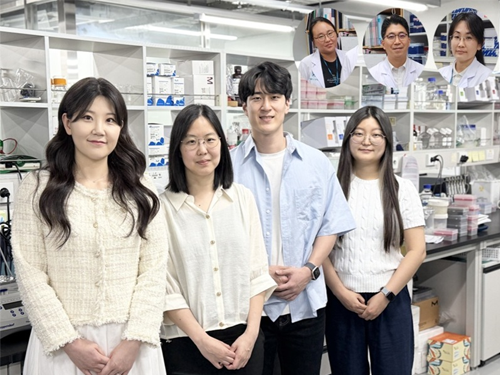 KAIST Develops Novel Candidiasis Treatment Overcoming Side Effects and Resistance
<(From left) Ph. D Candidate Ju Yeon Chung, Prof.Hyun Jung Chung, Ph.D candidate Seungju Yang, Ph.D candidate Ayoung Park, Dr. Yoon-Kyoung Hong from Asan Medical Center, Prof. Yong Pil Chong, Dr. Eunhee Jeon>
Candida, a type of fungus, which can spread throughout the body via the bloodstream, leading to organ damage and sepsis. Recently, the incidence of candidiasis has surged due to the increase in immunosuppressive therapies, medical implants, and transplantation. Korean researchers have successfully developed a next-generation treatment that, unlike existing antifungals, selectively acts only on Candida, achieving both high therapeutic efficacy and low side effects simultaneously.
KAIST (President Kwang Hyung Lee) announced on the 8th that a research team led by Professor Hyun-Jung Chung of the Department of Biological Sciences, in collaboration with Professor Yong Pil Jeong's team at Asan Medical Center, developed a gene-based nanotherapy (FTNx) that simultaneously inhibits two key enzymes in the Candida cell wall.
Current antifungal drugs for Candida have low target selectivity, which can affect human cells. Furthermore, their therapeutic efficacy is gradually decreasing due to the emergence of new resistant strains. Especially for immunocompromised patients, the infection progresses rapidly and has a poor prognosis, making the development of new treatments to overcome the limitations of existing therapies urgent.
The developed treatment can be administered systemically, and by combining gene suppression technology with nanomaterial technology, it effectively overcomes the structural limitations of existing compound-based drugs and successfully achieves selective treatment against only Candida.
The research team created a gold nanoparticle-based complex loaded with short DNA fragments called antisense oligonucleotides (ASO), which simultaneously target two crucial enzymes—β-1,3-glucan synthase (FKS1) and chitin synthase (CHS3)—important for forming the cell wall of the Candida fungus.
By applying a surface coating technology that binds to a specific glycolipid structure (a structure combining sugar and fat) on the Candida cell wall, a targeted delivery device was implemented. This successfully achieved a precise targeting effect, ensuring the complex is not delivered to human cells at all but acts selectively only on Candida.
<Figure 1: Overview of antifungal therapy design and experimental approach>
This complex, after entering Candida cells, cleaves the mRNA produced by the FKS1 and CHS3 genes, thereby inhibiting translation and simultaneously blocking the synthesis of cell wall components β-1,3-glucan and chitin. As a result, the
Candida cell wall loses its structural stability and collapses, suppressing bacterial survival and proliferation.
In fact, experiments using a systemic candidiasis model in mice confirmed the therapeutic effect: a significant reduction in
Candida count in the organs, normalization of immune responses, and a notable increase in survival rates were observed in the treated group.
Professor Hyun-Jung Chung, who led the research, stated, "This study presents a method to overcome the issues of human toxicity and drug resistance spread with existing treatments, marking an important turning point by demonstrating the applicability of gene therapy for systemic infections". She added, "We plan to continue research on optimizing administration methods and verifying toxicity for future clinical application."
This research involved Ju Yeon Chung and Yoon-Kyoung Hong as co-first authors , and was published in the international journal 'Nature Communications' on July 1st.
Paper Title: Effective treatment of systemic candidiasis by synergistic targeting of cell wall synthesis
DOI: 10.1038/s41467-025-60684-7
This research was supported by the Ministry of Health and Welfare and the National Research Foundation of Korea.
2025.07.08 View 3796
KAIST Develops Novel Candidiasis Treatment Overcoming Side Effects and Resistance
<(From left) Ph. D Candidate Ju Yeon Chung, Prof.Hyun Jung Chung, Ph.D candidate Seungju Yang, Ph.D candidate Ayoung Park, Dr. Yoon-Kyoung Hong from Asan Medical Center, Prof. Yong Pil Chong, Dr. Eunhee Jeon>
Candida, a type of fungus, which can spread throughout the body via the bloodstream, leading to organ damage and sepsis. Recently, the incidence of candidiasis has surged due to the increase in immunosuppressive therapies, medical implants, and transplantation. Korean researchers have successfully developed a next-generation treatment that, unlike existing antifungals, selectively acts only on Candida, achieving both high therapeutic efficacy and low side effects simultaneously.
KAIST (President Kwang Hyung Lee) announced on the 8th that a research team led by Professor Hyun-Jung Chung of the Department of Biological Sciences, in collaboration with Professor Yong Pil Jeong's team at Asan Medical Center, developed a gene-based nanotherapy (FTNx) that simultaneously inhibits two key enzymes in the Candida cell wall.
Current antifungal drugs for Candida have low target selectivity, which can affect human cells. Furthermore, their therapeutic efficacy is gradually decreasing due to the emergence of new resistant strains. Especially for immunocompromised patients, the infection progresses rapidly and has a poor prognosis, making the development of new treatments to overcome the limitations of existing therapies urgent.
The developed treatment can be administered systemically, and by combining gene suppression technology with nanomaterial technology, it effectively overcomes the structural limitations of existing compound-based drugs and successfully achieves selective treatment against only Candida.
The research team created a gold nanoparticle-based complex loaded with short DNA fragments called antisense oligonucleotides (ASO), which simultaneously target two crucial enzymes—β-1,3-glucan synthase (FKS1) and chitin synthase (CHS3)—important for forming the cell wall of the Candida fungus.
By applying a surface coating technology that binds to a specific glycolipid structure (a structure combining sugar and fat) on the Candida cell wall, a targeted delivery device was implemented. This successfully achieved a precise targeting effect, ensuring the complex is not delivered to human cells at all but acts selectively only on Candida.
<Figure 1: Overview of antifungal therapy design and experimental approach>
This complex, after entering Candida cells, cleaves the mRNA produced by the FKS1 and CHS3 genes, thereby inhibiting translation and simultaneously blocking the synthesis of cell wall components β-1,3-glucan and chitin. As a result, the
Candida cell wall loses its structural stability and collapses, suppressing bacterial survival and proliferation.
In fact, experiments using a systemic candidiasis model in mice confirmed the therapeutic effect: a significant reduction in
Candida count in the organs, normalization of immune responses, and a notable increase in survival rates were observed in the treated group.
Professor Hyun-Jung Chung, who led the research, stated, "This study presents a method to overcome the issues of human toxicity and drug resistance spread with existing treatments, marking an important turning point by demonstrating the applicability of gene therapy for systemic infections". She added, "We plan to continue research on optimizing administration methods and verifying toxicity for future clinical application."
This research involved Ju Yeon Chung and Yoon-Kyoung Hong as co-first authors , and was published in the international journal 'Nature Communications' on July 1st.
Paper Title: Effective treatment of systemic candidiasis by synergistic targeting of cell wall synthesis
DOI: 10.1038/s41467-025-60684-7
This research was supported by the Ministry of Health and Welfare and the National Research Foundation of Korea.
2025.07.08 View 3796 -
 KAIST Enhances Immunotherapy for Difficult-to-Treat Brain Tumors with Gut Microbiota
< Photo 1.(From left) Prof. Heung Kyu Lee, Department of Biological Sciences,
and Dr. Hyeon Cheol Kim>
Advanced treatments, known as immunotherapies that activate T cells—our body's immune cells—to eliminate cancer cells, have shown limited efficacy as standalone therapies for glioblastoma, the most lethal form of brain tumor. This is due to their minimal response to glioblastoma and high resistance to treatment.
Now, a KAIST research team has now demonstrated a new therapeutic strategy that can enhance the efficacy of immunotherapy for brain tumors by utilizing gut microbes and their metabolites. This also opens up possibilities for developing microbiome-based immunotherapy supplements in the future.
KAIST (President Kwang Hyung Lee) announced on July 1 that a research team led by Professor Heung Kyu Lee of the Department of Biological Sciences discovered and demonstrated a method to significantly improve the efficiency of glioblastoma immunotherapy by focusing on changes in the gut microbial ecosystem.
The research team noted that as glioblastoma progresses, the concentration of ‘tryptophan’, an important amino acid in the gut, sharply decreases, leading to changes in the gut microbial ecosystem. They discovered that by supplementing tryptophan to restore microbial diversity, specific beneficial strains activate CD8 T cells (a type of immune cell) and induce their infiltration into tumor tissues. Through a mouse model of glioblastoma, the research team confirmed that tryptophan supplementation enhanced the response of cancer-attacking T cells (especially CD8 T cells), leading to their increased migration to tumor sites such as lymph nodes and the brain.
In this process, they also revealed that ‘Duncaniella dubosii’, a beneficial commensal bacterium present in the gut, plays a crucial role. This bacterium helped T cells effectively redistribute within the body, and survival rates significantly improved when used in combination with immunotherapy (anti-PD-1).
Furthermore, it was demonstrated that even when this commensal bacterium was administered alone to germ-free mice (mice without any commensal microbes), the survival rate for glioblastoma increased. This is because the bacterium utilizes tryptophan to regulate the gut environment, and the metabolites produced in this process strengthen the ability of CD8 T cells to attack cancer cells.
Professor Heung Kyu Lee explained, "This research is a meaningful achievement, showing that even in intractable brain tumors where immune checkpoint inhibitors had no effect, a combined strategy utilizing gut microbes can significantly enhance treatment response."
Dr. Hyeon Cheol Kim of KAIST (currently a postdoctoral researcher at the Institute for Biological Sciences) participated as the first author. The research findings were published online in Cell Reports, an international journal in the life sciences, on June 26.
This research was conducted as part of the Basic Research Program and Bio & Medical Technology Development Program supported by the Ministry of Science and ICT and the National Research Foundation of Korea.
※Paper Title: Gut microbiota dysbiosis induced by brain tumor modulates the efficacy of immunotherapy
※DOI: https://doi.org/10.1016/j.celrep.2025.115825
2025.07.02 View 7195
KAIST Enhances Immunotherapy for Difficult-to-Treat Brain Tumors with Gut Microbiota
< Photo 1.(From left) Prof. Heung Kyu Lee, Department of Biological Sciences,
and Dr. Hyeon Cheol Kim>
Advanced treatments, known as immunotherapies that activate T cells—our body's immune cells—to eliminate cancer cells, have shown limited efficacy as standalone therapies for glioblastoma, the most lethal form of brain tumor. This is due to their minimal response to glioblastoma and high resistance to treatment.
Now, a KAIST research team has now demonstrated a new therapeutic strategy that can enhance the efficacy of immunotherapy for brain tumors by utilizing gut microbes and their metabolites. This also opens up possibilities for developing microbiome-based immunotherapy supplements in the future.
KAIST (President Kwang Hyung Lee) announced on July 1 that a research team led by Professor Heung Kyu Lee of the Department of Biological Sciences discovered and demonstrated a method to significantly improve the efficiency of glioblastoma immunotherapy by focusing on changes in the gut microbial ecosystem.
The research team noted that as glioblastoma progresses, the concentration of ‘tryptophan’, an important amino acid in the gut, sharply decreases, leading to changes in the gut microbial ecosystem. They discovered that by supplementing tryptophan to restore microbial diversity, specific beneficial strains activate CD8 T cells (a type of immune cell) and induce their infiltration into tumor tissues. Through a mouse model of glioblastoma, the research team confirmed that tryptophan supplementation enhanced the response of cancer-attacking T cells (especially CD8 T cells), leading to their increased migration to tumor sites such as lymph nodes and the brain.
In this process, they also revealed that ‘Duncaniella dubosii’, a beneficial commensal bacterium present in the gut, plays a crucial role. This bacterium helped T cells effectively redistribute within the body, and survival rates significantly improved when used in combination with immunotherapy (anti-PD-1).
Furthermore, it was demonstrated that even when this commensal bacterium was administered alone to germ-free mice (mice without any commensal microbes), the survival rate for glioblastoma increased. This is because the bacterium utilizes tryptophan to regulate the gut environment, and the metabolites produced in this process strengthen the ability of CD8 T cells to attack cancer cells.
Professor Heung Kyu Lee explained, "This research is a meaningful achievement, showing that even in intractable brain tumors where immune checkpoint inhibitors had no effect, a combined strategy utilizing gut microbes can significantly enhance treatment response."
Dr. Hyeon Cheol Kim of KAIST (currently a postdoctoral researcher at the Institute for Biological Sciences) participated as the first author. The research findings were published online in Cell Reports, an international journal in the life sciences, on June 26.
This research was conducted as part of the Basic Research Program and Bio & Medical Technology Development Program supported by the Ministry of Science and ICT and the National Research Foundation of Korea.
※Paper Title: Gut microbiota dysbiosis induced by brain tumor modulates the efficacy of immunotherapy
※DOI: https://doi.org/10.1016/j.celrep.2025.115825
2025.07.02 View 7195 -
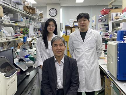 KAIST Identifies Master Regulator Blocking Immunotherapy, Paving the Way for a New Lung Cancer Treatment
Immune checkpoint inhibitors, a class of immunotherapies that help immune cells attack cancer more effectively, have revolutionized cancer treatment. However, fewer than 20% of patients respond to these treatments, highlighting the urgent need for new strategies tailored to both responders and non-responders.
KAIST researchers have discovered that 'DEAD-box helicases 54 (DDX54)', a type of RNA-binding protein, is the master regulator that hinders the effectiveness of immunotherapy—opening a new path for lung cancer treatment. This breakthrough technology has been transferred to faculty startup BioRevert Inc., where it is currently being developed as a companion therapeutic and is expected to enter clinical trials by 2028.
< Photo 1. (From left) Researcher Jungeun Lee, Professor Kwang-Hyun Cho and Postdoctoral Researcher Jeong-Ryeol Gong of the Department of Bio and Brain Engineering at KAIST >
KAIST (represented by President Kwang-Hyung Lee) announced on April 8 that a research team led by Professor Kwang-Hyun Cho from the Department of Bio and Brain Engineering had identified DDX54 as a critical factor that determines the immune evasion capacity of lung cancer cells. They demonstrated that suppressing DDX54 enhances immune cell infiltration into tumors and significantly improves the efficacy of immunotherapy.
Immunotherapy using anti-PD-1 or anti-PD-L1 antibodies is considered a powerful approach in cancer treatment. However, its low response rate limits the number of patients who actually benefit.
To identify likely responders, tumor mutational burden (TMB) has recently been approved by the FDA as a key biomarker for immunotherapy. Cancers with high mutation rates are thought to be more responsive to immune checkpoint inhibitors. However, even tumors with high TMB can display an “immune-desert” phenotype—where immune cell infiltration is severely limited—resulting in poor treatment responses.
< Figure 1. DDX54 was identified as the master regulator that induces resistance to immunotherapy by orchestrating suppression of immune cell infiltration through cancer tissues as lung cancer cells become immune-evasive >
Professor Kwang-Hyun Cho's research team compared transcriptome and genome data of lung cancer patients with immune evasion capabilities through gene regulatory network analysis (A) and discovered DDX54, a master regulator that induces resistance to immunotherapy (B-F).
This study is especially significant in that it successfully demonstrated that suppressing DDX54 in immune-desert lung tumors can overcome immunotherapy resistance and improve treatment outcomes.
The team used transcriptomic and genomic data from immune-evasive lung cancer patients and employed systems biology techniques to infer gene regulatory networks. Through this analysis, they identified DDX54 as a central regulator in the immune evasion of lung cancer cells.
In a syngeneic mouse model, the suppression of DDX54 led to significant increases in the infiltration of anti-cancer immune cells such as T cells and NK cells, and greatly improved the response to immunotherapy.
Single-cell transcriptomic and spatial transcriptomic analyses further showed that combination therapy targeting DDX54 promoted the differentiation of T cells and memory T cells that suppress tumors, while reducing the infiltration of regulatory T cells and exhausted T cells that support tumor growth.
< Figure 2. In the syngeneic mouse model made of lung cancer cells, it was confirmed that inhibiting DDX54 reversed the immune-evasion ability of cancer cells and enhanced the sensitivity to anti-PD-1 therapy >
In a syngeneic mouse model made of lung cancer cells exhibiting immunotherapy resistance, the treatment applied after DDX54 inhibition resulted in statistically significant inhibition of lung cancer growth (B-D) and a significant increase in immune cell infiltration into the tumor tissue (E, F).
The mechanism is believed to involve DDX54 suppression inactivating signaling pathways such as JAK-STAT, MYC, and NF-κB, thereby downregulating immune-evasive proteins CD38 and CD47. This also reduced the infiltration of circulating monocytes—which promote tumor development—and promoted the differentiation of M1 macrophages that play anti-tumor roles.
Professor Kwang-Hyun Cho stated, “We have, for the first time, identified a master regulatory factor that enables immune evasion in lung cancer cells. By targeting this factor, we developed a new therapeutic strategy that can induce responsiveness to immunotherapy in previously resistant cancers.”
He added, “The discovery of DDX54—hidden within the complex molecular networks of cancer cells—was made possible through the systematic integration of systems biology, combining IT and BT.”
The study, led by Professor Kwang-Hyun Cho, was published in the Proceedings of the National Academy of Sciences of the United States of America (PNAS) on April 2, 2025, with Jeong-Ryeol Gong being the first author, Jungeun Lee, a co-first author, and Younghyun Han, a co-author of the article.
< Figure 3. Single-cell transcriptome and spatial transcriptome analysis confirmed that knockdown of DDX54 increased immune cell infiltration into cancer tissues >
In a syngeneic mouse model made of lung cancer cells that underwent immunotherapy in combination with DDX54 inhibition, single-cell transcriptome (H-L) and spatial transcriptome (A-G) analysis of immune cells infiltrating inside cancer tissues were performed. As a result, it was confirmed that anticancer immune cells such as T cells, B cells, and NK cells actively infiltrated the core of lung cancer tissues when DDX54 inhibition and immunotherapy were concurrently administered.
(Paper title: “DDX54 downregulation enhances anti-PD1 therapy in immune-desert lung tumors with high tumor mutational burden,” DOI: https://doi.org/10.1073/pnas.2412310122)
This work was supported by the Ministry of Science and ICT and the National Research Foundation of Korea through the Mid-Career Research Program and Basic Research Laboratory Program.
< Figure 4. The identified master regulator DDX54 was confirmed to induce CD38 and CD47 expression through Jak-Stat3, MYC, and NF-κB activation. >
DDX54 activates the Jak-Stat3, MYC, and NF-κB pathways in lung cancer cells to increase CD38 and CD47 expression (A-G). This creates a cancer microenvironment that contributes to cancer development (H) and ultimately induces immune anticancer treatment resistance.
< Figure 5. It was confirmed that an immune-inflamed environment can be created by combining DDX54 inhibition and immune checkpoint inhibitor (ICI) therapy. >
When DDX54 inhibition and ICI therapy are simultaneously administered, the cancer cell characteristics change, the immune evasion ability is restored, and the environment is transformed into an ‘immune-activated’ environment in which immune cells easily infiltrate cancer tissues. This strengthens the anticancer immune response, thereby increasing the sensitivity of immunotherapy even in lung cancer tissues that previously had low responsiveness to immunotherapy.
2025.04.08 View 14367
KAIST Identifies Master Regulator Blocking Immunotherapy, Paving the Way for a New Lung Cancer Treatment
Immune checkpoint inhibitors, a class of immunotherapies that help immune cells attack cancer more effectively, have revolutionized cancer treatment. However, fewer than 20% of patients respond to these treatments, highlighting the urgent need for new strategies tailored to both responders and non-responders.
KAIST researchers have discovered that 'DEAD-box helicases 54 (DDX54)', a type of RNA-binding protein, is the master regulator that hinders the effectiveness of immunotherapy—opening a new path for lung cancer treatment. This breakthrough technology has been transferred to faculty startup BioRevert Inc., where it is currently being developed as a companion therapeutic and is expected to enter clinical trials by 2028.
< Photo 1. (From left) Researcher Jungeun Lee, Professor Kwang-Hyun Cho and Postdoctoral Researcher Jeong-Ryeol Gong of the Department of Bio and Brain Engineering at KAIST >
KAIST (represented by President Kwang-Hyung Lee) announced on April 8 that a research team led by Professor Kwang-Hyun Cho from the Department of Bio and Brain Engineering had identified DDX54 as a critical factor that determines the immune evasion capacity of lung cancer cells. They demonstrated that suppressing DDX54 enhances immune cell infiltration into tumors and significantly improves the efficacy of immunotherapy.
Immunotherapy using anti-PD-1 or anti-PD-L1 antibodies is considered a powerful approach in cancer treatment. However, its low response rate limits the number of patients who actually benefit.
To identify likely responders, tumor mutational burden (TMB) has recently been approved by the FDA as a key biomarker for immunotherapy. Cancers with high mutation rates are thought to be more responsive to immune checkpoint inhibitors. However, even tumors with high TMB can display an “immune-desert” phenotype—where immune cell infiltration is severely limited—resulting in poor treatment responses.
< Figure 1. DDX54 was identified as the master regulator that induces resistance to immunotherapy by orchestrating suppression of immune cell infiltration through cancer tissues as lung cancer cells become immune-evasive >
Professor Kwang-Hyun Cho's research team compared transcriptome and genome data of lung cancer patients with immune evasion capabilities through gene regulatory network analysis (A) and discovered DDX54, a master regulator that induces resistance to immunotherapy (B-F).
This study is especially significant in that it successfully demonstrated that suppressing DDX54 in immune-desert lung tumors can overcome immunotherapy resistance and improve treatment outcomes.
The team used transcriptomic and genomic data from immune-evasive lung cancer patients and employed systems biology techniques to infer gene regulatory networks. Through this analysis, they identified DDX54 as a central regulator in the immune evasion of lung cancer cells.
In a syngeneic mouse model, the suppression of DDX54 led to significant increases in the infiltration of anti-cancer immune cells such as T cells and NK cells, and greatly improved the response to immunotherapy.
Single-cell transcriptomic and spatial transcriptomic analyses further showed that combination therapy targeting DDX54 promoted the differentiation of T cells and memory T cells that suppress tumors, while reducing the infiltration of regulatory T cells and exhausted T cells that support tumor growth.
< Figure 2. In the syngeneic mouse model made of lung cancer cells, it was confirmed that inhibiting DDX54 reversed the immune-evasion ability of cancer cells and enhanced the sensitivity to anti-PD-1 therapy >
In a syngeneic mouse model made of lung cancer cells exhibiting immunotherapy resistance, the treatment applied after DDX54 inhibition resulted in statistically significant inhibition of lung cancer growth (B-D) and a significant increase in immune cell infiltration into the tumor tissue (E, F).
The mechanism is believed to involve DDX54 suppression inactivating signaling pathways such as JAK-STAT, MYC, and NF-κB, thereby downregulating immune-evasive proteins CD38 and CD47. This also reduced the infiltration of circulating monocytes—which promote tumor development—and promoted the differentiation of M1 macrophages that play anti-tumor roles.
Professor Kwang-Hyun Cho stated, “We have, for the first time, identified a master regulatory factor that enables immune evasion in lung cancer cells. By targeting this factor, we developed a new therapeutic strategy that can induce responsiveness to immunotherapy in previously resistant cancers.”
He added, “The discovery of DDX54—hidden within the complex molecular networks of cancer cells—was made possible through the systematic integration of systems biology, combining IT and BT.”
The study, led by Professor Kwang-Hyun Cho, was published in the Proceedings of the National Academy of Sciences of the United States of America (PNAS) on April 2, 2025, with Jeong-Ryeol Gong being the first author, Jungeun Lee, a co-first author, and Younghyun Han, a co-author of the article.
< Figure 3. Single-cell transcriptome and spatial transcriptome analysis confirmed that knockdown of DDX54 increased immune cell infiltration into cancer tissues >
In a syngeneic mouse model made of lung cancer cells that underwent immunotherapy in combination with DDX54 inhibition, single-cell transcriptome (H-L) and spatial transcriptome (A-G) analysis of immune cells infiltrating inside cancer tissues were performed. As a result, it was confirmed that anticancer immune cells such as T cells, B cells, and NK cells actively infiltrated the core of lung cancer tissues when DDX54 inhibition and immunotherapy were concurrently administered.
(Paper title: “DDX54 downregulation enhances anti-PD1 therapy in immune-desert lung tumors with high tumor mutational burden,” DOI: https://doi.org/10.1073/pnas.2412310122)
This work was supported by the Ministry of Science and ICT and the National Research Foundation of Korea through the Mid-Career Research Program and Basic Research Laboratory Program.
< Figure 4. The identified master regulator DDX54 was confirmed to induce CD38 and CD47 expression through Jak-Stat3, MYC, and NF-κB activation. >
DDX54 activates the Jak-Stat3, MYC, and NF-κB pathways in lung cancer cells to increase CD38 and CD47 expression (A-G). This creates a cancer microenvironment that contributes to cancer development (H) and ultimately induces immune anticancer treatment resistance.
< Figure 5. It was confirmed that an immune-inflamed environment can be created by combining DDX54 inhibition and immune checkpoint inhibitor (ICI) therapy. >
When DDX54 inhibition and ICI therapy are simultaneously administered, the cancer cell characteristics change, the immune evasion ability is restored, and the environment is transformed into an ‘immune-activated’ environment in which immune cells easily infiltrate cancer tissues. This strengthens the anticancer immune response, thereby increasing the sensitivity of immunotherapy even in lung cancer tissues that previously had low responsiveness to immunotherapy.
2025.04.08 View 14367