%62%69%6f%63%6f%6d%70%61%74%69%62%6c%65
-
 3D Stem Cell Culture Technology to Shift the Paradigm of Regenerative Medicine
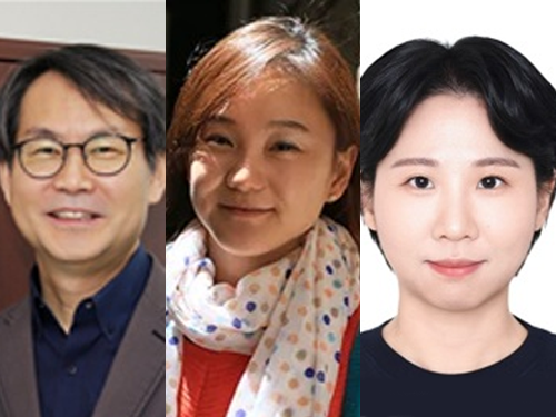
< (From left) KAIST Dr. Changjin Seo, Professor Sangyong Jon >
A breakthrough technology has been developed to overcome the limitation where stem cells fail to survive for long periods in the body, even when administered in large quantities. Stem cells are vital for regenerating damaged tissues or recovering injured areas. A KAIST research team has successfully enhanced both the survival rate and therapeutic efficacy of these cells by developing a 3D culture technology that precisely designs the cellular microenvironment. This achievement is expected to transcend the current limits of stem cell therapy and reshape the landscape of regenerative medicine.
On April 29th, the research team—led by Professor Sangyong Jon from the Department of Biological Sciences and featuring researchers Changjin Seo, Dohyeon Kim, Junhyuk Song, Sun-Young Kim, Youngju Son, and Afia Tasnim Rahman—announced the development of a novel culture technology to grow healthier stem cells. The team implemented a 3D platform by applying a polymer matrix (an artificial structure coating the culture substrate) to an "artificial floor" that mimics the natural in vivo environment. On this platform, they cultured human adipose-derived stem cells (hADSCs) in three dimensions, confirming a dramatic improvement in cellular function and therapeutic impact.
Human adipose-derived stem cells have been favored for clinical use due to their ease of harvest, high proliferation, and low immune rejection. However, traditional 2D (planar) culture methods cause cells to age and lose function over time. Previous 3D methods, such as forming cell aggregates (spheroids), also faced hurdles in maintaining long-term survival and functionality within the body.
To solve this, the research team developed a densely cross-linked synthetic polymer material composed of siloxane (a biocompatible polymer of silicon and oxygen), named "poly-Z."
This material modifies the physicochemical properties of the culture substrate to promote the adsorption of albumin proteins found in the culture medium. As a result, cells do not adhere to the floor but instead self-assemble into 3D spheroid structures. These spheroids showed increased production of the extracellular matrix (ECM), creating an environment highly similar to the human body and demonstrating performance far superior to conventional methods.
Experimental results showed that stem cells cultured on the poly-Z platform exhibited enhanced differentiation potential and immunomodulatory functions, with a significantly increased survival time inside the body.
< Schematic of hADSC Spheroid Formation on the Synthetic Polymer Matrix, Poly-Z >
Notably, in animal models of acute colitis and acute liver injury, this method showed significantly higher therapeutic efficacy than conventional methods. This suggests that even with the same dosage, the cells live longer and act more vigorously. The team confirmed that the activation of integrin and FAK signaling pathways—the mechanisms through which cells sense and respond to their environment—strengthened the stem cells' functions, allowing them to better perceive their surroundings and perform more effectively after transplantation.
Professor Sangyong Jon stated, "This research proves that a precisely engineered synthetic polymer-based 3D environment can simultaneously enhance the function and therapeutic efficacy of stem cells. We expect this to be widely utilized in developing next-generation cell therapies for various incurable diseases, including inflammatory conditions."
The study, with Dr. Changjin Seo from the KAIST InnoCORE AI-Drug Discovery Center as the lead author, was published online on March 31 in the international journal Advanced Science (Impact Factor: 14.1).
Paper Title: Polymer Matrix-Based 3D Culture Significantly Enhances the Differentiation and Immunomodulatory Functions of Human Adipose-Derived Stem Cells
DOI: https://doi.org/10.1002/advs.202518704
This research was supported by the Korea Multi-Ministry Regenerative Medicine Project, the KAIST InnoCORE Program, and the Leader Research Grant of the National Research Foundation of Korea.
2026.04.29 View 284
3D Stem Cell Culture Technology to Shift the Paradigm of Regenerative Medicine
< (From left) KAIST Dr. Changjin Seo, Professor Sangyong Jon >
A breakthrough technology has been developed to overcome the limitation where stem cells fail to survive for long periods in the body, even when administered in large quantities. Stem cells are vital for regenerating damaged tissues or recovering injured areas. A KAIST research team has successfully enhanced both the survival rate and therapeutic efficacy of these cells by developing a 3D culture technology that precisely designs the cellular microenvironment. This achievement is expected to transcend the current limits of stem cell therapy and reshape the landscape of regenerative medicine.
On April 29th, the research team—led by Professor Sangyong Jon from the Department of Biological Sciences and featuring researchers Changjin Seo, Dohyeon Kim, Junhyuk Song, Sun-Young Kim, Youngju Son, and Afia Tasnim Rahman—announced the development of a novel culture technology to grow healthier stem cells. The team implemented a 3D platform by applying a polymer matrix (an artificial structure coating the culture substrate) to an "artificial floor" that mimics the natural in vivo environment. On this platform, they cultured human adipose-derived stem cells (hADSCs) in three dimensions, confirming a dramatic improvement in cellular function and therapeutic impact.
Human adipose-derived stem cells have been favored for clinical use due to their ease of harvest, high proliferation, and low immune rejection. However, traditional 2D (planar) culture methods cause cells to age and lose function over time. Previous 3D methods, such as forming cell aggregates (spheroids), also faced hurdles in maintaining long-term survival and functionality within the body.
To solve this, the research team developed a densely cross-linked synthetic polymer material composed of siloxane (a biocompatible polymer of silicon and oxygen), named "poly-Z."
This material modifies the physicochemical properties of the culture substrate to promote the adsorption of albumin proteins found in the culture medium. As a result, cells do not adhere to the floor but instead self-assemble into 3D spheroid structures. These spheroids showed increased production of the extracellular matrix (ECM), creating an environment highly similar to the human body and demonstrating performance far superior to conventional methods.
Experimental results showed that stem cells cultured on the poly-Z platform exhibited enhanced differentiation potential and immunomodulatory functions, with a significantly increased survival time inside the body.
< Schematic of hADSC Spheroid Formation on the Synthetic Polymer Matrix, Poly-Z >
Notably, in animal models of acute colitis and acute liver injury, this method showed significantly higher therapeutic efficacy than conventional methods. This suggests that even with the same dosage, the cells live longer and act more vigorously. The team confirmed that the activation of integrin and FAK signaling pathways—the mechanisms through which cells sense and respond to their environment—strengthened the stem cells' functions, allowing them to better perceive their surroundings and perform more effectively after transplantation.
Professor Sangyong Jon stated, "This research proves that a precisely engineered synthetic polymer-based 3D environment can simultaneously enhance the function and therapeutic efficacy of stem cells. We expect this to be widely utilized in developing next-generation cell therapies for various incurable diseases, including inflammatory conditions."
The study, with Dr. Changjin Seo from the KAIST InnoCORE AI-Drug Discovery Center as the lead author, was published online on March 31 in the international journal Advanced Science (Impact Factor: 14.1).
Paper Title: Polymer Matrix-Based 3D Culture Significantly Enhances the Differentiation and Immunomodulatory Functions of Human Adipose-Derived Stem Cells
DOI: https://doi.org/10.1002/advs.202518704
This research was supported by the Korea Multi-Ministry Regenerative Medicine Project, the KAIST InnoCORE Program, and the Leader Research Grant of the National Research Foundation of Korea.
2026.04.29 View 284 -
 Development of Low-Frequency Wireless Sensor for Real-Time Monitoring of Arteriosclerosis Without Electromagnetic Interference Concerns
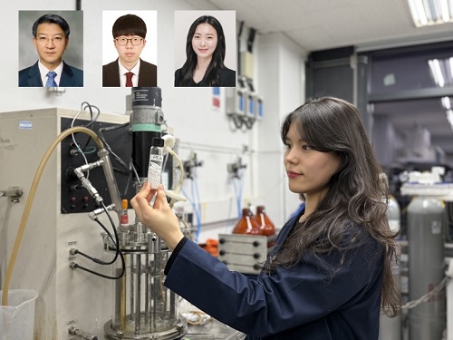
< (Top, left to right) Dr. Haerim Kim (KAIST), Ph.D. candidate Ji Hong Kim (Hanyang University), student Jaewon Rhee (KAIST); (Bottom, left to right) Prof. Seungyoung Ahn (KAIST), Prof. Do Hwan Kim (Hanyang University) >
Wireless sensors used in wearable smart devices and medical equipment must be capable of detecting minute changes while maintaining high operational stability. However, existing technologies often utilize excessively high frequencies, leading to electromagnetic interference (EMI) or potential health risks to the human body. To address these fundamental issues, a Korean research team has developed a low-frequency-based wireless sensor technology.
A joint research team, led by Professor Seungyoung Ahn from the KAIST Cho Chun Shik Graduate School of Mobility and Professor Do Hwan Kim from the Department of Chemical Engineering at Hanyang University, announced the development of "WiLECS" (Wireless Ionic-Electronic Coupling System), a low-frequency wireless electrochemical sensing platform that combines ion-based materials with wireless power transfer technology.
Conventional wireless sensors suffer from low capacitance (the ability to store electrical charge), requiring high frequencies in the megahertz (MHz) range to compensate. However, these high-frequency methods can cause tissue heating or signal instability, limiting their practical application in clinical medical settings.
To solve this, the Hanyang University team developed a biocompatible ionic material with high capacitance, leveraging the movement of ions to store significant amounts of electricity. The KAIST team then integrated this with a wireless LC resonance system—a circuit that exchanges energy wirelessly. The result is a wireless sensor that operates stably within the human body at low frequencies.
Specifically, the team designed the system such that ions are attached to the surface of gold nanoparticles, inhibiting their movement under normal conditions and releasing them only when pressure is applied. This design causes a significant change in electrical storage even under minor stimuli. By monitoring these changes through fluctuations in the wireless frequency, the sensor can detect extremely subtle variations in pressure. The system demonstrates excellent performance even in the sub-1 MHz frequency band and achieves a high Signal-to-Noise Ratio (SNR) due to reduced electromagnetic interference.
In experiments using an artificial blood vessel model, the research team successfully monitored real-time blood pressure changes associated with arteriosclerosis—a condition where blood vessels harden or narrow. This demonstrates the technology's strong potential for future cardiovascular disease monitoring.
< Schematic Diagram of a Wireless Blood Pressure Monitoring Platform (AI-Generated Image) >
This study is significant as it shifts away from the conventional approach of simply increasing frequency to improve performance. Instead, it solves the problem by fundamentally altering the physical mechanism of sensor operation. It is evaluated as opening a new path for the design of next-generation bio-devices where electromagnetic safety is paramount.
Professor Seungyoung Ahn stated, "This research is a result of a collaborative effort combining ionic materials and wireless technology, overcoming the limitations of existing high-frequency wireless sensors. It has great potential for expansion as a platform that enables stable wireless sensing while minimizing electromagnetic impact."
The study, with Haerim Kim (KAIST) and Ji Hong Kim (Hanyang University) as joint first authors, was published in the world-renowned academic journal Nature Communications on March 11.
Paper Title: Low-frequency ionic-electronic coupling for energy-efficient noise-resilient wireless bioelectronics
DOI: https://www.nature.com/articles/s41467-026-70331-4
Authors: Ji Hong Kim (Hanyang University, Co-first author), Haerim Kim (KAIST, Co-first author), Jaewon Rhee (KAIST, Co-author), Joo Sung Kim, Hanbin Choi, Won Hyuk Choi, Yoseph Park, Jong Hwi Kim, So Young Kim, Seungyoung Ahn (KAIST, Corresponding author), and Do Hwan Kim (Hanyang University, Corresponding author).
2026.04.13 View 384
Development of Low-Frequency Wireless Sensor for Real-Time Monitoring of Arteriosclerosis Without Electromagnetic Interference Concerns
< (Top, left to right) Dr. Haerim Kim (KAIST), Ph.D. candidate Ji Hong Kim (Hanyang University), student Jaewon Rhee (KAIST); (Bottom, left to right) Prof. Seungyoung Ahn (KAIST), Prof. Do Hwan Kim (Hanyang University) >
Wireless sensors used in wearable smart devices and medical equipment must be capable of detecting minute changes while maintaining high operational stability. However, existing technologies often utilize excessively high frequencies, leading to electromagnetic interference (EMI) or potential health risks to the human body. To address these fundamental issues, a Korean research team has developed a low-frequency-based wireless sensor technology.
A joint research team, led by Professor Seungyoung Ahn from the KAIST Cho Chun Shik Graduate School of Mobility and Professor Do Hwan Kim from the Department of Chemical Engineering at Hanyang University, announced the development of "WiLECS" (Wireless Ionic-Electronic Coupling System), a low-frequency wireless electrochemical sensing platform that combines ion-based materials with wireless power transfer technology.
Conventional wireless sensors suffer from low capacitance (the ability to store electrical charge), requiring high frequencies in the megahertz (MHz) range to compensate. However, these high-frequency methods can cause tissue heating or signal instability, limiting their practical application in clinical medical settings.
To solve this, the Hanyang University team developed a biocompatible ionic material with high capacitance, leveraging the movement of ions to store significant amounts of electricity. The KAIST team then integrated this with a wireless LC resonance system—a circuit that exchanges energy wirelessly. The result is a wireless sensor that operates stably within the human body at low frequencies.
Specifically, the team designed the system such that ions are attached to the surface of gold nanoparticles, inhibiting their movement under normal conditions and releasing them only when pressure is applied. This design causes a significant change in electrical storage even under minor stimuli. By monitoring these changes through fluctuations in the wireless frequency, the sensor can detect extremely subtle variations in pressure. The system demonstrates excellent performance even in the sub-1 MHz frequency band and achieves a high Signal-to-Noise Ratio (SNR) due to reduced electromagnetic interference.
In experiments using an artificial blood vessel model, the research team successfully monitored real-time blood pressure changes associated with arteriosclerosis—a condition where blood vessels harden or narrow. This demonstrates the technology's strong potential for future cardiovascular disease monitoring.
< Schematic Diagram of a Wireless Blood Pressure Monitoring Platform (AI-Generated Image) >
This study is significant as it shifts away from the conventional approach of simply increasing frequency to improve performance. Instead, it solves the problem by fundamentally altering the physical mechanism of sensor operation. It is evaluated as opening a new path for the design of next-generation bio-devices where electromagnetic safety is paramount.
Professor Seungyoung Ahn stated, "This research is a result of a collaborative effort combining ionic materials and wireless technology, overcoming the limitations of existing high-frequency wireless sensors. It has great potential for expansion as a platform that enables stable wireless sensing while minimizing electromagnetic impact."
The study, with Haerim Kim (KAIST) and Ji Hong Kim (Hanyang University) as joint first authors, was published in the world-renowned academic journal Nature Communications on March 11.
Paper Title: Low-frequency ionic-electronic coupling for energy-efficient noise-resilient wireless bioelectronics
DOI: https://www.nature.com/articles/s41467-026-70331-4
Authors: Ji Hong Kim (Hanyang University, Co-first author), Haerim Kim (KAIST, Co-first author), Jaewon Rhee (KAIST, Co-author), Joo Sung Kim, Hanbin Choi, Won Hyuk Choi, Yoseph Park, Jong Hwi Kim, So Young Kim, Seungyoung Ahn (KAIST, Corresponding author), and Do Hwan Kim (Hanyang University, Corresponding author).
2026.04.13 View 384 -
 KAIST Researchers Unveil Technical Principles Behind Antibacterial Graphene Toothbrushes with 10 Million Units Sold
< (From left) Professor Hyun Jung Chung , Ph.D candidate Ju Yeon Chung, Ph.D candidate Sujin Cha, Professor Sang Ouk Kim >
Hygiene in everyday items that touch the body—such as clothing, masks, and toothbrushes—is critically important. The underlying principle of how graphene selectively eliminates only bacteria has now been revealed. A KAIST research team has presented the potential for a next-generation antibacterial material that is safe for the human body and capable of replacing antibiotics.
KAIST announced on March 25th that a joint research team, led by Professor Sang Ouk Kim from the Department of Materials Science and Engineering and Professor Hyun Jung Chung from the Department of Biological Sciences, has identified the mechanism by which Graphene Oxide (GO) exhibits powerful antibacterial effects against bacteria while remaining harmless to human cells. Graphene oxide is a nanomaterial consisting of an atomic level carbon layer (graphene) with oxygen attached; it is characterized by its ability to mix well with water and implement various functions.
This study is highly significant as it provides molecular-level proof of graphene's antibacterial action, which had not been clearly understood until now.
The research team confirmed that graphene oxide performs "selective antibacterial action" by attaching to and destroying only the membranes of bacteria, much like a magnet attaches only to specific metals, while leaving human cells untouched. This occurs because the oxygen functional groups on the surface of graphene oxide selectively bind with a specific component (POPG) found only in bacterial cell membranes. Simply put, it recognizes a "target" present only in bacterial membranes to attach and destroy the structure. In this context, phospholipids are fatty components that make up the membrane surrounding a cell, and POPG is a component primarily present in bacteria.
< Schematic diagram of the selective interaction between graphene oxide and cell membranes >
< Identification of selective interaction mechanisms at the molecular level through microscopic and chemical analysis of artificial lipid vesicles mimicking cell membranes >
Nanofibers applying this principle effectively inhibited the growth of various pathogenic bacteria, including superbugs resistant to antibiotics. Animal experiments also confirmed its effectiveness in promoting wound healing without inducing inflammation.
< Verification of antibacterial and wound healing enhancement effects in a porcine infected wound model >
Furthermore, fibers using this material maintained their antibacterial functions even after multiple washes, showing potential for use in various industrial fields such as apparel and medical textiles.
This technology is already being applied to consumer products. The graphene antibacterial toothbrush, released through the original patents of the faculty-led startup 'Materials Creation Co., Ltd.,' has sold over 10 million units, proving its commercial viability. Additionally, GrapheneTex—textile materiala incorporating this technology—was used in the uniforms of the Taekwondo demonstration team at the 2024 Paris Olympics and is expected to play an active role in functional sportswear at upcoming international sporting events like the 2026 Asian Games.
< Commercially available graphene toothbrush >
< Graphene material image (AI-generated image) >
Professor Sang Ouk Kim explained, "This study is an example of scientifically uncovering why graphene can selectively kill bacteria while remaining safe for the human body." He emphasized, "By utilizing this principle, we can expand beyond safe clothing without harsh chemicals to an infinite range of applications, including wearable devices and medical textile systems."
Sujin Cha (PhD program, Department of Materials Science and Engineering) and Ju Yeon Chung (Integrated MS/PhD program, Department of Biological Sciences) participated as first authors. Professor Hyun Jung Chung participated as a co-corresponding author. The research was published on March 2nd in the prestigious materials science journal, Advanced Functional Materials.
※ Paper Title: Biocompatible but Antibacterial Mechanism of Graphene Oxide for Sustainable Antibiotics, DOI: 10.1002/adfm.202313583
Additionally, Nanowerk (http://www.nanowerk.com/), a global portal for nanotechnology, featured these findings as a 'Spotlight' titled "Graphene oxide destroys bacteria without harming human tissue."
This research was conducted with support from the 'Nano/Material Technology Development (R&D)' program, the 'Individual Basic Research' program, and the 'Mid-Career Researcher Support Program' funded by the Ministry of Science and ICT.
2026.03.26 View 3946
KAIST Researchers Unveil Technical Principles Behind Antibacterial Graphene Toothbrushes with 10 Million Units Sold
< (From left) Professor Hyun Jung Chung , Ph.D candidate Ju Yeon Chung, Ph.D candidate Sujin Cha, Professor Sang Ouk Kim >
Hygiene in everyday items that touch the body—such as clothing, masks, and toothbrushes—is critically important. The underlying principle of how graphene selectively eliminates only bacteria has now been revealed. A KAIST research team has presented the potential for a next-generation antibacterial material that is safe for the human body and capable of replacing antibiotics.
KAIST announced on March 25th that a joint research team, led by Professor Sang Ouk Kim from the Department of Materials Science and Engineering and Professor Hyun Jung Chung from the Department of Biological Sciences, has identified the mechanism by which Graphene Oxide (GO) exhibits powerful antibacterial effects against bacteria while remaining harmless to human cells. Graphene oxide is a nanomaterial consisting of an atomic level carbon layer (graphene) with oxygen attached; it is characterized by its ability to mix well with water and implement various functions.
This study is highly significant as it provides molecular-level proof of graphene's antibacterial action, which had not been clearly understood until now.
The research team confirmed that graphene oxide performs "selective antibacterial action" by attaching to and destroying only the membranes of bacteria, much like a magnet attaches only to specific metals, while leaving human cells untouched. This occurs because the oxygen functional groups on the surface of graphene oxide selectively bind with a specific component (POPG) found only in bacterial cell membranes. Simply put, it recognizes a "target" present only in bacterial membranes to attach and destroy the structure. In this context, phospholipids are fatty components that make up the membrane surrounding a cell, and POPG is a component primarily present in bacteria.
< Schematic diagram of the selective interaction between graphene oxide and cell membranes >
< Identification of selective interaction mechanisms at the molecular level through microscopic and chemical analysis of artificial lipid vesicles mimicking cell membranes >
Nanofibers applying this principle effectively inhibited the growth of various pathogenic bacteria, including superbugs resistant to antibiotics. Animal experiments also confirmed its effectiveness in promoting wound healing without inducing inflammation.
< Verification of antibacterial and wound healing enhancement effects in a porcine infected wound model >
Furthermore, fibers using this material maintained their antibacterial functions even after multiple washes, showing potential for use in various industrial fields such as apparel and medical textiles.
This technology is already being applied to consumer products. The graphene antibacterial toothbrush, released through the original patents of the faculty-led startup 'Materials Creation Co., Ltd.,' has sold over 10 million units, proving its commercial viability. Additionally, GrapheneTex—textile materiala incorporating this technology—was used in the uniforms of the Taekwondo demonstration team at the 2024 Paris Olympics and is expected to play an active role in functional sportswear at upcoming international sporting events like the 2026 Asian Games.
< Commercially available graphene toothbrush >
< Graphene material image (AI-generated image) >
Professor Sang Ouk Kim explained, "This study is an example of scientifically uncovering why graphene can selectively kill bacteria while remaining safe for the human body." He emphasized, "By utilizing this principle, we can expand beyond safe clothing without harsh chemicals to an infinite range of applications, including wearable devices and medical textile systems."
Sujin Cha (PhD program, Department of Materials Science and Engineering) and Ju Yeon Chung (Integrated MS/PhD program, Department of Biological Sciences) participated as first authors. Professor Hyun Jung Chung participated as a co-corresponding author. The research was published on March 2nd in the prestigious materials science journal, Advanced Functional Materials.
※ Paper Title: Biocompatible but Antibacterial Mechanism of Graphene Oxide for Sustainable Antibiotics, DOI: 10.1002/adfm.202313583
Additionally, Nanowerk (http://www.nanowerk.com/), a global portal for nanotechnology, featured these findings as a 'Spotlight' titled "Graphene oxide destroys bacteria without harming human tissue."
This research was conducted with support from the 'Nano/Material Technology Development (R&D)' program, the 'Individual Basic Research' program, and the 'Mid-Career Researcher Support Program' funded by the Ministry of Science and ICT.
2026.03.26 View 3946 -
 Hemostasis in 1 Second... Boosting Survival Rates for Soldiers
< (From top left) Professor Steve Park, Professor Sangyong Jon, (From bottom left) President Kwang-Hyung Lee, Ph.D canddiate Youngju Son, Ph.D candidate Kyusoon Park >
The leading cause of death due to injuries in war is excessive bleeding. A KAIST research team, in which an Army Major participated, has tackled this issue head-on. By developing a next-generation powder-type hemostatic agent that stops bleeding in one second just by spraying it, they have presented an innovative technology that will change the paradigm of combatant survivability.
KAIST announced on December 29th that a joint research team led by Professor Steve Park from the Department of Materials Science and Engineering and Professor Sangyong Jon from the Department of Biological Sciences has developed a powder-type hemostatic agent that forms a powerful hydrogel barrier within approximately one second when sprayed on a wound.
This technology reached a high level of perfection as a practical technology considering real combat environments, with an Army Major researcher directly participating in the study. By implementing characteristics that allow instant hardening even under extreme conditions such as combat and disaster sites due to high usability and storage stability, immediate emergency treatment is possible.
Until now, patch-type hemostatic agents widely used in medical fields have had limitations in application to deep and complex wounds due to their flat structure, and were sensitive to temperature and humidity, posing limits on storage and operation.
Accordingly, the research team developed a next-generation hemostatic agent in powder form that can be freely applied even to deep, large, and irregular wounds. They have secured versatility to respond to various types of wounds with a single powder.
< AGCL powder development strategy and fabrication schematin/ Gelation speed and blood absorption capacity of AGCL powder >
Existing powder hemostatic agents had limits in hemostatic capability as they functioned by physically absorbing blood to form a barrier. To solve this problem, the research team focused on the ionic reactions within the blood.
The ‘AGCL powder’ developed this time has a structure that combines biocompatible natural materials such as Alginate and Gellan Gum (which react with calcium for ultra-fast gelation and physical sealing) and Chitosan (which bonds with blood components to enhance chemical and biological hemostasis). It reacts with cations such as calcium in the blood to turn into a gel state in one second, instantly sealing the wound.
Furthermore, by forming a three-dimensional structure inside the powder, it can absorb blood amounting to more than 7 times its own weight (725%). Due to this, it quickly blocks blood flow even in high-pressure and excessive bleeding situations, and showed superior sealing performance compared to commercial hemostatic agents with a high adhesive strength of over '40kPa', a level of pressure that can withstand being pressed strongly by hand.
AGCL powder is composed entirely of naturally derived materials, showing a hemolysis rate of less than 3%, a cell viability rate of over 99%, and an antibacterial effect of 99.9%, making it safe even when in contact with blood. In animal experiments, excellent tissue regeneration effects such as rapid wound recovery and promotion of blood vessel and collagen regeneration were confirmed.
In surgical liver injury experiments, the amount of bleeding and hemostasis time were significantly reduced compared to commercial hemostatic agents, and liver function recovered to normal levels two weeks after surgery. No abnormal findings were observed in systemic toxicity evaluations.
In particular, this hemostatic agent maintains its performance for two years even in room temperature and high humidity environments, possessing the advantage of being ready for immediate use in harsh environments such as military operation sites or disaster areas.
Although this research is an advanced new material technology developed with national defense purposes in mind, it has great potential for application throughout emergency medicine, including disaster sites, developing countries, and medically underserved areas.
It is evaluated as a representative spin-off case* where national defense science and technology expanded to the private sector, as it is capable of everything from emergency treatment on the battlefield to internal surgical hemostasis.
*Spin-off case: Expanding or transferring national defense science and technology for use in the private sector. Examples include computers, GPS, microwave ovens, etc.
< Validation of efficacy in wounds through animal experiments / Validation of efficacy in a liver surgery model >
This study was recognized for its scientific innovation and national defense utility simultaneously, winning the 2025 KAIST Q-Day President's Award and the Minister of National Defense Award at the 2024 KAIST-KNDU National Defense Academic Conference.
Ph.D candidate Kyusoon Park (Army Major), who participated in the research, stated, “The core of modern warfare is minimizing the loss of human life,” and added, “I started the research with a sense of mission to save even one more soldier.” He continued, “I hope this technology will be used as a life-saving technology in both national defense and private medical fields.”
This research, in which KAIST PhD student Kyusoon Park and Ph.D candidate Youngju Son participated as lead authors and was guided by Professor Steve Park and Professor Sangyong Jon, was published online on October 28, 2025, in the international academic journal in the field of chemistry/materials engineering, Advanced Functional Materials (IF 19.0).
※ Paper Title: An Ionic Gelation Powder for Ultrafast Hemostasis and Accelerated Wound Healing, DOI: 10.1002/adfm.202523910
Meanwhile, this research was conducted with the support of the National Research Foundation of Korea (NRF)."
2025.12.29 View 13594
Hemostasis in 1 Second... Boosting Survival Rates for Soldiers
< (From top left) Professor Steve Park, Professor Sangyong Jon, (From bottom left) President Kwang-Hyung Lee, Ph.D canddiate Youngju Son, Ph.D candidate Kyusoon Park >
The leading cause of death due to injuries in war is excessive bleeding. A KAIST research team, in which an Army Major participated, has tackled this issue head-on. By developing a next-generation powder-type hemostatic agent that stops bleeding in one second just by spraying it, they have presented an innovative technology that will change the paradigm of combatant survivability.
KAIST announced on December 29th that a joint research team led by Professor Steve Park from the Department of Materials Science and Engineering and Professor Sangyong Jon from the Department of Biological Sciences has developed a powder-type hemostatic agent that forms a powerful hydrogel barrier within approximately one second when sprayed on a wound.
This technology reached a high level of perfection as a practical technology considering real combat environments, with an Army Major researcher directly participating in the study. By implementing characteristics that allow instant hardening even under extreme conditions such as combat and disaster sites due to high usability and storage stability, immediate emergency treatment is possible.
Until now, patch-type hemostatic agents widely used in medical fields have had limitations in application to deep and complex wounds due to their flat structure, and were sensitive to temperature and humidity, posing limits on storage and operation.
Accordingly, the research team developed a next-generation hemostatic agent in powder form that can be freely applied even to deep, large, and irregular wounds. They have secured versatility to respond to various types of wounds with a single powder.
< AGCL powder development strategy and fabrication schematin/ Gelation speed and blood absorption capacity of AGCL powder >
Existing powder hemostatic agents had limits in hemostatic capability as they functioned by physically absorbing blood to form a barrier. To solve this problem, the research team focused on the ionic reactions within the blood.
The ‘AGCL powder’ developed this time has a structure that combines biocompatible natural materials such as Alginate and Gellan Gum (which react with calcium for ultra-fast gelation and physical sealing) and Chitosan (which bonds with blood components to enhance chemical and biological hemostasis). It reacts with cations such as calcium in the blood to turn into a gel state in one second, instantly sealing the wound.
Furthermore, by forming a three-dimensional structure inside the powder, it can absorb blood amounting to more than 7 times its own weight (725%). Due to this, it quickly blocks blood flow even in high-pressure and excessive bleeding situations, and showed superior sealing performance compared to commercial hemostatic agents with a high adhesive strength of over '40kPa', a level of pressure that can withstand being pressed strongly by hand.
AGCL powder is composed entirely of naturally derived materials, showing a hemolysis rate of less than 3%, a cell viability rate of over 99%, and an antibacterial effect of 99.9%, making it safe even when in contact with blood. In animal experiments, excellent tissue regeneration effects such as rapid wound recovery and promotion of blood vessel and collagen regeneration were confirmed.
In surgical liver injury experiments, the amount of bleeding and hemostasis time were significantly reduced compared to commercial hemostatic agents, and liver function recovered to normal levels two weeks after surgery. No abnormal findings were observed in systemic toxicity evaluations.
In particular, this hemostatic agent maintains its performance for two years even in room temperature and high humidity environments, possessing the advantage of being ready for immediate use in harsh environments such as military operation sites or disaster areas.
Although this research is an advanced new material technology developed with national defense purposes in mind, it has great potential for application throughout emergency medicine, including disaster sites, developing countries, and medically underserved areas.
It is evaluated as a representative spin-off case* where national defense science and technology expanded to the private sector, as it is capable of everything from emergency treatment on the battlefield to internal surgical hemostasis.
*Spin-off case: Expanding or transferring national defense science and technology for use in the private sector. Examples include computers, GPS, microwave ovens, etc.
< Validation of efficacy in wounds through animal experiments / Validation of efficacy in a liver surgery model >
This study was recognized for its scientific innovation and national defense utility simultaneously, winning the 2025 KAIST Q-Day President's Award and the Minister of National Defense Award at the 2024 KAIST-KNDU National Defense Academic Conference.
Ph.D candidate Kyusoon Park (Army Major), who participated in the research, stated, “The core of modern warfare is minimizing the loss of human life,” and added, “I started the research with a sense of mission to save even one more soldier.” He continued, “I hope this technology will be used as a life-saving technology in both national defense and private medical fields.”
This research, in which KAIST PhD student Kyusoon Park and Ph.D candidate Youngju Son participated as lead authors and was guided by Professor Steve Park and Professor Sangyong Jon, was published online on October 28, 2025, in the international academic journal in the field of chemistry/materials engineering, Advanced Functional Materials (IF 19.0).
※ Paper Title: An Ionic Gelation Powder for Ultrafast Hemostasis and Accelerated Wound Healing, DOI: 10.1002/adfm.202523910
Meanwhile, this research was conducted with the support of the National Research Foundation of Korea (NRF)."
2025.12.29 View 13594 -
 Jaewook Myung, First Korean Selected as '40 Under 40 Recognition Program' Next Generation Environmental Engineering Leader
< Professor Jaewook Myung of KAIST Department of Civil and Environmental Engineering >
KAIST announced on December 12th that Professor Jaewook Myung of the Department of Civil and Environmental Engineering was selected as the first Korean recipient of the '40 Under 40 Recognition Program' for Next Generation Environmental Engineering Leaders, organized by the American Academy of Environmental Engineers and Scientists (AAEES).
< The '40 Under 40 Recognition Program' is an international award program selecting next-generation leaders in the field of Environmental Engineering and Science >
This award is presented annually by AAEES to select next-generation environmental engineering researchers who demonstrate innovative research achievements, social contribution, and educational leadership. Professor Myung's selection is particularly significant as he is the first Korean to be chosen since the program's inception. The award ceremony is scheduled to be held in Washington D.C. in April 2026.
AAEES is the world's highest-authority professional organization leading the global environmental engineering sector through operating the Professional Environmental Engineer (PEE) certification system, policy consultation, and international academic exchange. This award is highly regarded for greatly enhancing the international standing of domestic environmental engineering and sustainability research.
Amid the deepening problems of plastic waste increase and greenhouse gas emissions, where existing technologies are showing limitations in providing solutions, Professor Jaewook Myung has garnered significant attention from academia and industry by developing technology to convert greenhouse gases such as methane ($CH_4$) and carbon dioxide ($CO_2$) into biodegradable plastics. His research is highly praised for presenting a new industrial paradigm that fuses environmental microbiology and materials science to convert greenhouse gases into high-value bio-materials.
Professor Myung's research team secured microbial metabolic control technology to transform greenhouse gases into materials, an accelerated process that simultaneously enhances the synthesis and decomposition efficiency of plastics, and pilot process design and engineering technology applicable in industrial settings. This established a sustainable circular technology model capable of simultaneously addressing greenhouse gas reduction and plastic pollution issues.
Furthermore, the research team expanded these foundational technologies to develop various application products, such as biodegradable coating materials that naturally decompose in the ocean, biocompatible bio-based electronic materials, and industrial 3D printing filaments, realizing full-cycle innovation from basic research to application and industrialization. These achievements are recognized as world-class sustainable technology alternatives that can simultaneously overcome the problems of plastic downcycling and the economic limitations of greenhouse gas utilization technology.
Professor Myung also shows excellent performance in nurturing talent. His advised students are growing into next-generation environmental and sustainability researchers, having won major awards both domestically and internationally, including the American Chemical Society (ACS) Environmental Chemistry Graduate Student Award, the Presidential Science Scholarship, the Merck Innovation Cup Prize, and the Republic of Korea Talent Award. He is also establishing himself as a leading researcher in the commercialization of sustainable technology by expanding his research achievements into the social and industrial ecosystem through technology collaboration with industries, patents, and consultation with public institutions.
The AAEES Selection Committee evaluated Professor Jaewook Myung as "a researcher possessing technical excellence, social responsibility, and educational leadership, and an innovator who has pioneered new areas of environmental engineering." Professor Myung expressed his thoughts, saying, "This award is a result made possible by the students who researched and challenged alongside me and the collaborative research culture of KAIST," and added, "I will contribute to brightening the future of humanity and the planet through sustainable resource circulation technology."
2025.12.12 View 2010
Jaewook Myung, First Korean Selected as '40 Under 40 Recognition Program' Next Generation Environmental Engineering Leader
< Professor Jaewook Myung of KAIST Department of Civil and Environmental Engineering >
KAIST announced on December 12th that Professor Jaewook Myung of the Department of Civil and Environmental Engineering was selected as the first Korean recipient of the '40 Under 40 Recognition Program' for Next Generation Environmental Engineering Leaders, organized by the American Academy of Environmental Engineers and Scientists (AAEES).
< The '40 Under 40 Recognition Program' is an international award program selecting next-generation leaders in the field of Environmental Engineering and Science >
This award is presented annually by AAEES to select next-generation environmental engineering researchers who demonstrate innovative research achievements, social contribution, and educational leadership. Professor Myung's selection is particularly significant as he is the first Korean to be chosen since the program's inception. The award ceremony is scheduled to be held in Washington D.C. in April 2026.
AAEES is the world's highest-authority professional organization leading the global environmental engineering sector through operating the Professional Environmental Engineer (PEE) certification system, policy consultation, and international academic exchange. This award is highly regarded for greatly enhancing the international standing of domestic environmental engineering and sustainability research.
Amid the deepening problems of plastic waste increase and greenhouse gas emissions, where existing technologies are showing limitations in providing solutions, Professor Jaewook Myung has garnered significant attention from academia and industry by developing technology to convert greenhouse gases such as methane ($CH_4$) and carbon dioxide ($CO_2$) into biodegradable plastics. His research is highly praised for presenting a new industrial paradigm that fuses environmental microbiology and materials science to convert greenhouse gases into high-value bio-materials.
Professor Myung's research team secured microbial metabolic control technology to transform greenhouse gases into materials, an accelerated process that simultaneously enhances the synthesis and decomposition efficiency of plastics, and pilot process design and engineering technology applicable in industrial settings. This established a sustainable circular technology model capable of simultaneously addressing greenhouse gas reduction and plastic pollution issues.
Furthermore, the research team expanded these foundational technologies to develop various application products, such as biodegradable coating materials that naturally decompose in the ocean, biocompatible bio-based electronic materials, and industrial 3D printing filaments, realizing full-cycle innovation from basic research to application and industrialization. These achievements are recognized as world-class sustainable technology alternatives that can simultaneously overcome the problems of plastic downcycling and the economic limitations of greenhouse gas utilization technology.
Professor Myung also shows excellent performance in nurturing talent. His advised students are growing into next-generation environmental and sustainability researchers, having won major awards both domestically and internationally, including the American Chemical Society (ACS) Environmental Chemistry Graduate Student Award, the Presidential Science Scholarship, the Merck Innovation Cup Prize, and the Republic of Korea Talent Award. He is also establishing himself as a leading researcher in the commercialization of sustainable technology by expanding his research achievements into the social and industrial ecosystem through technology collaboration with industries, patents, and consultation with public institutions.
The AAEES Selection Committee evaluated Professor Jaewook Myung as "a researcher possessing technical excellence, social responsibility, and educational leadership, and an innovator who has pioneered new areas of environmental engineering." Professor Myung expressed his thoughts, saying, "This award is a result made possible by the students who researched and challenged alongside me and the collaborative research culture of KAIST," and added, "I will contribute to brightening the future of humanity and the planet through sustainable resource circulation technology."
2025.12.12 View 2010 -
 KAIST Presents Game-Changing Technology for Intractable Brain Disease Treatment Using Micro OLEDs
<(From left)Professor Kyung Cheol Choi, Professor Hyunjoo J. Lee, Dr. Somin Lee from the School of Electrical Engineering>
Optogenetics is a technique that controls neural activity by stimulating neurons expressing light-sensitive proteins with specific wavelengths of light. It has opened new possibilities for identifying causes of brain disorders and developing treatments for intractable neurological diseases. Because this technology requires precise stimulation inside the human brain with minimal damage to soft brain tissue, it must be integrated into a neural probe—a medical device implanted in the brain. KAIST researchers have now proposed a new paradigm for neural probes by integrating micro OLEDs into thin, flexible, implantable medical devices.
KAIST (President Kwang Hyung Lee) announced on the 6th of July that professor Kyung Cheol Choi and professor Hyunjoo J. Lee from the School of Electrical Engineering have jointly succeeded in developing an optogenetic neural probe integrated with flexible micro OLEDs.
Optical fibers have been used for decades in optogenetic research to deliver light to deep brain regions from external light sources. Recently, research has focused on flexible optical fibers and ultra-miniaturized neural probes that integrate light sources for single-neuron stimulation.
The research team focused on micro OLEDs due to their high spatial resolution and flexibility, which allow for precise light delivery to small areas of neurons. This enables detailed brain circuit analysis while minimizing side effects and avoiding restrictions on animal movement. Moreover, micro OLEDs offer precise control of light wavelengths and support multi-site stimulation, making them suitable for studying complex brain functions.
However, the device's electrical properties degrade easily in the presence of moisture or water, which limited their use as implantable bioelectronics. Furthermore, optimizing the high-resolution integration process on thin, flexible probes remained a challenge.
To address this, the team enhanced the operational reliability of OLEDs in moist, oxygen-rich environments and minimized tissue damage during implantation. They patterned an ultrathin, flexible encapsulation layer* composed of aluminum oxide and parylene-C (Al₂O₃/parylene-C) at widths of 260–600 micrometers (μm) to maintain biocompatibility.
*Encapsulation layer: A barrier that completely blocks oxygen and water molecules from the external environment, ensuring the longevity and reliability of the device.
When integrating the high-resolution micro OLEDs, the researchers also used parylene-C, the same biocompatible material as the encapsulation layer, to maintain flexibility and safety. To eliminate electrical interference between adjacent OLED pixels and spatially separate them, they introduced a pixel define layer (PDL), enabling the independent operation of eight micro OLEDs.
Furthermore, they precisely controlled the residual stress and thickness in the multilayer film structure of the device, ensuring its flexibility even in biological environments. This optimization allowed for probe insertion without bending or external shuttles or needles, minimizing mechanical stress during implantation.
2025.07.07 View 5547
KAIST Presents Game-Changing Technology for Intractable Brain Disease Treatment Using Micro OLEDs
<(From left)Professor Kyung Cheol Choi, Professor Hyunjoo J. Lee, Dr. Somin Lee from the School of Electrical Engineering>
Optogenetics is a technique that controls neural activity by stimulating neurons expressing light-sensitive proteins with specific wavelengths of light. It has opened new possibilities for identifying causes of brain disorders and developing treatments for intractable neurological diseases. Because this technology requires precise stimulation inside the human brain with minimal damage to soft brain tissue, it must be integrated into a neural probe—a medical device implanted in the brain. KAIST researchers have now proposed a new paradigm for neural probes by integrating micro OLEDs into thin, flexible, implantable medical devices.
KAIST (President Kwang Hyung Lee) announced on the 6th of July that professor Kyung Cheol Choi and professor Hyunjoo J. Lee from the School of Electrical Engineering have jointly succeeded in developing an optogenetic neural probe integrated with flexible micro OLEDs.
Optical fibers have been used for decades in optogenetic research to deliver light to deep brain regions from external light sources. Recently, research has focused on flexible optical fibers and ultra-miniaturized neural probes that integrate light sources for single-neuron stimulation.
The research team focused on micro OLEDs due to their high spatial resolution and flexibility, which allow for precise light delivery to small areas of neurons. This enables detailed brain circuit analysis while minimizing side effects and avoiding restrictions on animal movement. Moreover, micro OLEDs offer precise control of light wavelengths and support multi-site stimulation, making them suitable for studying complex brain functions.
However, the device's electrical properties degrade easily in the presence of moisture or water, which limited their use as implantable bioelectronics. Furthermore, optimizing the high-resolution integration process on thin, flexible probes remained a challenge.
To address this, the team enhanced the operational reliability of OLEDs in moist, oxygen-rich environments and minimized tissue damage during implantation. They patterned an ultrathin, flexible encapsulation layer* composed of aluminum oxide and parylene-C (Al₂O₃/parylene-C) at widths of 260–600 micrometers (μm) to maintain biocompatibility.
*Encapsulation layer: A barrier that completely blocks oxygen and water molecules from the external environment, ensuring the longevity and reliability of the device.
When integrating the high-resolution micro OLEDs, the researchers also used parylene-C, the same biocompatible material as the encapsulation layer, to maintain flexibility and safety. To eliminate electrical interference between adjacent OLED pixels and spatially separate them, they introduced a pixel define layer (PDL), enabling the independent operation of eight micro OLEDs.
Furthermore, they precisely controlled the residual stress and thickness in the multilayer film structure of the device, ensuring its flexibility even in biological environments. This optimization allowed for probe insertion without bending or external shuttles or needles, minimizing mechanical stress during implantation.
2025.07.07 View 5547 -
 KAIST Develops Eco-Friendly, Nylon-Like Plastic Using Microorganisms
Poly(ester amide) amide is a next-generation material that combines the advantages of PET (polyester) and nylon (polyamide), two widely used plastics. However, it could only be produced from fossil fuels, which posed environmental concerns. Using microorganisms, KAIST researchers have successfully developed a new bio-based plastic to replace conventional plastic.
KAIST (represented by President Kwang Hyung Lee) announced on the 20th of March that a research team led by Distinguished Professor Sang Yup Lee from the Department of Chemical and Biomolecular Engineering has developed microbial strains through systems metabolic engineering to produce various eco-friendly, bio-based poly(ester amide)s. The team collaborated with researchers from the Korea Research Institute of Chemical Technology (KRICT, President Young-Kook Lee) to analyze and confirm the properties of the resulting plastic.
Professor Sang Yup Lee’s research team designed new metabolic pathways that do not naturally exist in microorganisms, and developed a platform microbial strain capable of producing nine different types of poly(ester amide)s, including poly(3-hydroxybutyrate-ran-3-aminopropionate) and poly(3-hydroxybutyrate-ran-4-aminobutyrate).
Using glucose derived from abundant biomass sources such as waste wood and weeds, the team successfully produced poly(ester amide)s in an eco-friendly manner. The researchers also confirmed the potential for industrial-scale production by demonstrating high production efficiency (54.57 g/L) using fed-batch fermentation of the engineered strain.
In collaboration with researchers Haemin Jeong and Jihoon Shin from KRICT, the KAIST team analyzed the properties of the bio-based plastic and found that it exhibited characteristics similar to high-density polyethylene (HDPE). This means the new plastic is not only eco-friendly but also strong and durable enough to replace conventional plastics.
The engineered strains and strategies developed in this study are expected to be useful not only for producing various poly(ester amide)s but also for constructing metabolic pathways for the biosynthesis of other types of polymers.
Professor Sang Yup Lee stated, “This study is the first to demonstrate the possibility of producing poly(ester amide)s (plastics) through a renewable bio-based chemical process rather than relying on the petroleum-based chemical industry. We plan to further enhance the production yield and efficiency through continued research.”
The study was published online on March 17 in the international journal Nature Chemical Biology.
·Title: Biosynthesis of poly(ester amide)s in engineered Escherichia coli
·DOI: 10.1038/s41589-025-01842-2
·Authors: A total of seven authors including Tong Un Chae (KAIST, first author), So Young Choi (KAIST, second author), Da-Hee Ahn (KAIST, third author), Woo Dae Jang (KAIST, fourth author), Haemin Jeong (KRICT, fifth author), Jihoon Shin (KRICT, sixth author), and Sang Yup Lee (KAIST, corresponding author).
This research was supported by the Ministry of Science and ICT (MSIT) under the Eco-Friendly Chemical Technology Development Project as part of the "Next-Generation Biorefinery Technology Development to Lead the Bio-Chemical Industry" initiative (project led by Distinguished Professor Sang Yup Lee at KAIST).
2025.03.24 View 12739
KAIST Develops Eco-Friendly, Nylon-Like Plastic Using Microorganisms
Poly(ester amide) amide is a next-generation material that combines the advantages of PET (polyester) and nylon (polyamide), two widely used plastics. However, it could only be produced from fossil fuels, which posed environmental concerns. Using microorganisms, KAIST researchers have successfully developed a new bio-based plastic to replace conventional plastic.
KAIST (represented by President Kwang Hyung Lee) announced on the 20th of March that a research team led by Distinguished Professor Sang Yup Lee from the Department of Chemical and Biomolecular Engineering has developed microbial strains through systems metabolic engineering to produce various eco-friendly, bio-based poly(ester amide)s. The team collaborated with researchers from the Korea Research Institute of Chemical Technology (KRICT, President Young-Kook Lee) to analyze and confirm the properties of the resulting plastic.
Professor Sang Yup Lee’s research team designed new metabolic pathways that do not naturally exist in microorganisms, and developed a platform microbial strain capable of producing nine different types of poly(ester amide)s, including poly(3-hydroxybutyrate-ran-3-aminopropionate) and poly(3-hydroxybutyrate-ran-4-aminobutyrate).
Using glucose derived from abundant biomass sources such as waste wood and weeds, the team successfully produced poly(ester amide)s in an eco-friendly manner. The researchers also confirmed the potential for industrial-scale production by demonstrating high production efficiency (54.57 g/L) using fed-batch fermentation of the engineered strain.
In collaboration with researchers Haemin Jeong and Jihoon Shin from KRICT, the KAIST team analyzed the properties of the bio-based plastic and found that it exhibited characteristics similar to high-density polyethylene (HDPE). This means the new plastic is not only eco-friendly but also strong and durable enough to replace conventional plastics.
The engineered strains and strategies developed in this study are expected to be useful not only for producing various poly(ester amide)s but also for constructing metabolic pathways for the biosynthesis of other types of polymers.
Professor Sang Yup Lee stated, “This study is the first to demonstrate the possibility of producing poly(ester amide)s (plastics) through a renewable bio-based chemical process rather than relying on the petroleum-based chemical industry. We plan to further enhance the production yield and efficiency through continued research.”
The study was published online on March 17 in the international journal Nature Chemical Biology.
·Title: Biosynthesis of poly(ester amide)s in engineered Escherichia coli
·DOI: 10.1038/s41589-025-01842-2
·Authors: A total of seven authors including Tong Un Chae (KAIST, first author), So Young Choi (KAIST, second author), Da-Hee Ahn (KAIST, third author), Woo Dae Jang (KAIST, fourth author), Haemin Jeong (KRICT, fifth author), Jihoon Shin (KRICT, sixth author), and Sang Yup Lee (KAIST, corresponding author).
This research was supported by the Ministry of Science and ICT (MSIT) under the Eco-Friendly Chemical Technology Development Project as part of the "Next-Generation Biorefinery Technology Development to Lead the Bio-Chemical Industry" initiative (project led by Distinguished Professor Sang Yup Lee at KAIST).
2025.03.24 View 12739 -
 Novel High-performance and Sustainable Paper Coating Material created by KAIST-Yonsei University Research Team to reduce microplastic pollution
What if there is a biodegradable packaging material with high performance without leaving microplastics?
Plastic pollution presents a global challenge that must be solved. In particular, packaging accounts for 30-50% of the total plastic consumption. While paper packaging is eco-friendly, it lacks crucial functionalities like moisture resistance and strength. Traditional coating materials exacerbate plastic pollution, prompting the need for sustainable alternatives.
Polyethylene (PE) and ethylene vinyl alcohol (EVOH) are typically used as coating materials to improve the low barrier properties of paper packaging, but these substances do not decompose and worsen microplastic pollution when disposed of in the natural environment. In response to this problem, packaging materials made from bio-based substances and biodegradable plastics have been developed, but in most cases, as the packaging performance improves, the biodegradability diminishes rapidly.
KAIST announced that a joint research team led by Professor Jaewook Myung of the Department of Civil and Environmental Engineering, Professor Hanseul Yang of the Department of Life Sciences, and Professor Jongcheol Seo of the Department of Packaging and Logistics <Figure 4. Back cover art of Green Chemistry journal of the latest volume, describing the boric acid cross-linked poly(vinyl alcohol) coated paper featuring marine biodegradability, biocompatibility, high barrier properties, and robustness developed through this study.>
at Yonsei University tackled the challenge of balancing packaging performance and sustainability. They successfully developed a sustainable, marine biodegradable, high-performance paper coating material.
* Biodegradable plastic: A plastic that can be decomposed by microorganisms in natural environments such as soil and ocean or artificial conditions such as industrial composting and anaerobic digestion by microorganisms.
*Microplastics: Tiny pieces of plastic less than 5 mm, produced during the decomposition of bulk plastic materials. Microplastics can persist in the sea for more than decades, causing severe marine pollution.
The team utilized boric acid-crosslinked poly(vinyl alcohol) (PVA), a biodegradable plastic, to coat the paper, thereby enhancing its biodegradability, barrier properties, and strength. The resulting coated paper exhibited superior performance compared to conventional plastics, with excellent barrier properties and physical strength, even in humid conditions.
<Figure 1. (a) Chemical structure of boric acid-crosslinked poly(vinyl alcohol) coating on paper, (b-c) Oxygen and water vapor barrier properties, (d-f) Tensile strength in dry and moist conditions. OTR: Oxygen transmission rate, WVTR: Water vapor transmission rate.>
The team also conducted an in-depth examination of biodegradation and biocompatibility to systematically evaluate the sustainability of the newly developed coated paper. Biodegradation was assessed by simulating the marine environment, known for its challenging biodegradability conditions. The team employed a respiratory system-based bioreactor to measure the degree of carbon mineralization into carbon dioxide. After 111 days of biodegradation, it was found that the coated papers achieved 59-82% biodegradation depending on the coating component. The phenomenon in which marine bacteria are decomposing the coating material was captured through a scanning electron microscope. In addition, in vitro biocompatibility was confirmed through human embryonic kidney and mouse embryonic fibroblast cells, as well as high in-vivo biocompatibility of the coated paper was verified through mouse experiments.
Through this study, the joint research team proposed a coating strategy that can improve packaging performance while upholding sustainability to address the drawbacks of paper packaging. The boric acid-crosslinked PVA-coated paper eliminates the need for artificial composting conditions or sewage treatment facilities. Being biodegradable in natural environments and characterized by low toxicity, this newly coated paper does not exacerbate environmental pollution when accidentally discarded. Thus, it presents a sustainable substitute for plastic packaging materials.
<Figure 2. (a) Normal paper and boric acid-crosslinked poly(vinyl alcohol) coated paper, (b) Biodegradation of the coated paper by marine bacteria, (c) Result of cytotoxicity test using human embryonic kidney and mouse embryonic fibroblast cells. (d) Vital organs after one-month exposure of the coated papers to mice.>
Professor Jaewook Myung at KAIST, who led the sustainability study of coated paper, said, "The development of a marine biodegradable high-performance paper coating is the result of combining the innovative technologies of three leading research teams in each field." He said, “We will continue to develop sustainable materials with excellent performance.” Meanwhile, Professor Jongchul Seo of Yonsei University, who led the research on the development of high-performance paper coating, mentioned, “Through this research, we have developed a sustainable paper packaging technology that can replace non-degradable plastic packaging, and we expect the research outcome will be applied in industry,”.
<Figure 3. End-of-life scenario of papers coated by BA-crosslinked PVA in the marine environment. The coated papers potentially be disintegrated by marine microorganisms and ocean waves and tides. The depolymerization of PVA coating and paper is then mediated by extracellular depolymerases such as oxidases and cellulases, after which the small subunits (oligomers and monomers) are assimilated by microbial cells. The carbon components in the coated papers are ultimately mineralized into CO2, posing no harm in the ocean.>
The work was published in Green Chemistry and Food Chemistry journals. This study was conducted with the support of the Korea Research Foundation and the Korea Institute for Agriculture, Food and Rural Affairs Technology Planning and Evaluation, etc.
*Title of paper published in Green Chemistry: Boric acid-crosslinked poly(vinyl alcohol): biodegradable, biocompatible, robust, and high-barrier paper coating
※ Selected as the article for the back cover of the journal .
- Authors: Shinhyeong Choe, Seulki You, Kitae Park, Youngju Kim, Jehee Park, Yongjun Cho, Jongchul Seo, Hanseul Yang, and Jaewook Myung)
- Date: April 17, 2024
- DOI: 10.1039/D4GC00618F
*Title of paper published in Food Chemistry: Effect of epichlorohydrin treatment on the coating process and performance of high-barrier paper packaging
- Authors: Kitae Park, Shinhyeong Choe, Kambiz Sadeghi, Pradeep Kumar Panda, Jaewook Myung, Dowan Kim, and Jongchul Seo
- Date: February 19, 2024
- DOI: 10.1016/j.foodchem.2024.138772
<Figure 4. Back cover art of Green Chemistry journal of the latest volume, describing the boric acid cross-linked poly(vinyl alcohol) coated paper featuring marine biodegradability, biocompatibility, high barrier properties, and robustness developed through this study.>
2024.05.22 View 14447
Novel High-performance and Sustainable Paper Coating Material created by KAIST-Yonsei University Research Team to reduce microplastic pollution
What if there is a biodegradable packaging material with high performance without leaving microplastics?
Plastic pollution presents a global challenge that must be solved. In particular, packaging accounts for 30-50% of the total plastic consumption. While paper packaging is eco-friendly, it lacks crucial functionalities like moisture resistance and strength. Traditional coating materials exacerbate plastic pollution, prompting the need for sustainable alternatives.
Polyethylene (PE) and ethylene vinyl alcohol (EVOH) are typically used as coating materials to improve the low barrier properties of paper packaging, but these substances do not decompose and worsen microplastic pollution when disposed of in the natural environment. In response to this problem, packaging materials made from bio-based substances and biodegradable plastics have been developed, but in most cases, as the packaging performance improves, the biodegradability diminishes rapidly.
KAIST announced that a joint research team led by Professor Jaewook Myung of the Department of Civil and Environmental Engineering, Professor Hanseul Yang of the Department of Life Sciences, and Professor Jongcheol Seo of the Department of Packaging and Logistics <Figure 4. Back cover art of Green Chemistry journal of the latest volume, describing the boric acid cross-linked poly(vinyl alcohol) coated paper featuring marine biodegradability, biocompatibility, high barrier properties, and robustness developed through this study.>
at Yonsei University tackled the challenge of balancing packaging performance and sustainability. They successfully developed a sustainable, marine biodegradable, high-performance paper coating material.
* Biodegradable plastic: A plastic that can be decomposed by microorganisms in natural environments such as soil and ocean or artificial conditions such as industrial composting and anaerobic digestion by microorganisms.
*Microplastics: Tiny pieces of plastic less than 5 mm, produced during the decomposition of bulk plastic materials. Microplastics can persist in the sea for more than decades, causing severe marine pollution.
The team utilized boric acid-crosslinked poly(vinyl alcohol) (PVA), a biodegradable plastic, to coat the paper, thereby enhancing its biodegradability, barrier properties, and strength. The resulting coated paper exhibited superior performance compared to conventional plastics, with excellent barrier properties and physical strength, even in humid conditions.
<Figure 1. (a) Chemical structure of boric acid-crosslinked poly(vinyl alcohol) coating on paper, (b-c) Oxygen and water vapor barrier properties, (d-f) Tensile strength in dry and moist conditions. OTR: Oxygen transmission rate, WVTR: Water vapor transmission rate.>
The team also conducted an in-depth examination of biodegradation and biocompatibility to systematically evaluate the sustainability of the newly developed coated paper. Biodegradation was assessed by simulating the marine environment, known for its challenging biodegradability conditions. The team employed a respiratory system-based bioreactor to measure the degree of carbon mineralization into carbon dioxide. After 111 days of biodegradation, it was found that the coated papers achieved 59-82% biodegradation depending on the coating component. The phenomenon in which marine bacteria are decomposing the coating material was captured through a scanning electron microscope. In addition, in vitro biocompatibility was confirmed through human embryonic kidney and mouse embryonic fibroblast cells, as well as high in-vivo biocompatibility of the coated paper was verified through mouse experiments.
Through this study, the joint research team proposed a coating strategy that can improve packaging performance while upholding sustainability to address the drawbacks of paper packaging. The boric acid-crosslinked PVA-coated paper eliminates the need for artificial composting conditions or sewage treatment facilities. Being biodegradable in natural environments and characterized by low toxicity, this newly coated paper does not exacerbate environmental pollution when accidentally discarded. Thus, it presents a sustainable substitute for plastic packaging materials.
<Figure 2. (a) Normal paper and boric acid-crosslinked poly(vinyl alcohol) coated paper, (b) Biodegradation of the coated paper by marine bacteria, (c) Result of cytotoxicity test using human embryonic kidney and mouse embryonic fibroblast cells. (d) Vital organs after one-month exposure of the coated papers to mice.>
Professor Jaewook Myung at KAIST, who led the sustainability study of coated paper, said, "The development of a marine biodegradable high-performance paper coating is the result of combining the innovative technologies of three leading research teams in each field." He said, “We will continue to develop sustainable materials with excellent performance.” Meanwhile, Professor Jongchul Seo of Yonsei University, who led the research on the development of high-performance paper coating, mentioned, “Through this research, we have developed a sustainable paper packaging technology that can replace non-degradable plastic packaging, and we expect the research outcome will be applied in industry,”.
<Figure 3. End-of-life scenario of papers coated by BA-crosslinked PVA in the marine environment. The coated papers potentially be disintegrated by marine microorganisms and ocean waves and tides. The depolymerization of PVA coating and paper is then mediated by extracellular depolymerases such as oxidases and cellulases, after which the small subunits (oligomers and monomers) are assimilated by microbial cells. The carbon components in the coated papers are ultimately mineralized into CO2, posing no harm in the ocean.>
The work was published in Green Chemistry and Food Chemistry journals. This study was conducted with the support of the Korea Research Foundation and the Korea Institute for Agriculture, Food and Rural Affairs Technology Planning and Evaluation, etc.
*Title of paper published in Green Chemistry: Boric acid-crosslinked poly(vinyl alcohol): biodegradable, biocompatible, robust, and high-barrier paper coating
※ Selected as the article for the back cover of the journal .
- Authors: Shinhyeong Choe, Seulki You, Kitae Park, Youngju Kim, Jehee Park, Yongjun Cho, Jongchul Seo, Hanseul Yang, and Jaewook Myung)
- Date: April 17, 2024
- DOI: 10.1039/D4GC00618F
*Title of paper published in Food Chemistry: Effect of epichlorohydrin treatment on the coating process and performance of high-barrier paper packaging
- Authors: Kitae Park, Shinhyeong Choe, Kambiz Sadeghi, Pradeep Kumar Panda, Jaewook Myung, Dowan Kim, and Jongchul Seo
- Date: February 19, 2024
- DOI: 10.1016/j.foodchem.2024.138772
<Figure 4. Back cover art of Green Chemistry journal of the latest volume, describing the boric acid cross-linked poly(vinyl alcohol) coated paper featuring marine biodegradability, biocompatibility, high barrier properties, and robustness developed through this study.>
2024.05.22 View 14447 -
 A KAIST Research Team Develops a Novel “Bone Bandage” Material for Cracked Bones
Bone regeneration is a complex process, and existing methods to aid regeneration including transplants and growth factor transmissions face limitations such as the high cost. But recently, a piezoelectric material that can promote the growth of bone tissue has been developed.
A KAIST research team led by Professor Seungbum Hong from the Department of Materials Science and Engineering (DMSE) announced on January 25 the development of a biomimetic scaffold that generates electrical signals upon the application of pressure by utilizing the unique osteogenic ability of hydroxyapatite (HAp). This research was conducted in collaboration with a team led by Professor Jangho Kim from the Department of Convergence Biosystems Engineering at Chonnam National University.
HAp is a basic calcium phosphate material found in bones and teeth. This biocompatible mineral substance is also known to prevent tooth decay and is often used in toothpaste.
Previous studies on piezoelectric scaffolds confirmed the effects of piezoelectricity on promoting bone regeneration and improving bone fusion in various polymer-based materials, but were limited in simulating the complex cellular environment required for optimal bone tissue regeneration. However, this research suggests a new method for utilizing the unique osteogenic abilities of HAp to develop a material that mimics the environment for bone tissue in a living body.
< Figure 1. Design and characterization of piezoelectrically and topographically originated biomimetic scaffolds. (a) Schematic representation of the enhanced bone regeneration mechanism through electrical and topographical cues provided by HAp-incorporated P(VDF-TrFE) scaffolds. (b) Schematic diagram of the fabrication process. >
The research team developed a manufacturing process that fuses HAp with a polymer film. The flexible and free-standing scaffold developed through this process demonstrated its remarkable potential for promoting bone regeneration through in-vitro and in-vivo experiments in rats.
The team also identified the principles of bone regeneration that their scaffold is based on. Using atomic force microscopy (AFM), they analysed the electrical properties of the scaffold and evaluated the detailed surface properties related to cell shape and cell skeletal protein formation. They also investigated the effects of piezoelectricity and surface properties on the expression of growth factors.
Professor Hong from KAIST’s DMSE said, “We have developed a HAp-based piezoelectric composite material that can act like a ‘bone bandage’ through its ability to accelerate bone regeneration.” He added, “This research not only suggests a new direction for designing biomaterials, but is also significant in having explored the effects of piezoelectricity and surface properties on bone regeneration.”
This research, conducted by co-first authors Soyun Joo and Soyeon Kim from Professor Hong’s group, was published on ACS Applied Materials & Interfaces on January 4 under the title “Piezoelectrically and Topographically Engineered Scaffolds for Accelerating Bone Regeneration”. From Professor Kim’s group, Ph.D. candidate Yonghyun Gwon also participated as co-first author, and Professor Kim himself as a corresponding author.
< Figure 2. Analysis of piezoelectric and surface properties of the biomimetic scaffolds using atomic force microscopy. (a) PFM amplitude and phase images of box-poled composite scaffolds. The white bar represents 2 μm. (b) 3D representations of composite scaffolds paired with typical 2D line sections. (c) In vivo bone regeneration micro-CT analysis, (d) schematic representation of filler-derived electrical origins in bone regeneration. >
This research was supported by the KAIST Research and Development Team, the KUSTAR-KAIST Joint Research Center, the KAIST Global Singularity Project, and the government-funded Basic Research Project by the National Research Foundation of Korea.
2024.02.01 View 13292
A KAIST Research Team Develops a Novel “Bone Bandage” Material for Cracked Bones
Bone regeneration is a complex process, and existing methods to aid regeneration including transplants and growth factor transmissions face limitations such as the high cost. But recently, a piezoelectric material that can promote the growth of bone tissue has been developed.
A KAIST research team led by Professor Seungbum Hong from the Department of Materials Science and Engineering (DMSE) announced on January 25 the development of a biomimetic scaffold that generates electrical signals upon the application of pressure by utilizing the unique osteogenic ability of hydroxyapatite (HAp). This research was conducted in collaboration with a team led by Professor Jangho Kim from the Department of Convergence Biosystems Engineering at Chonnam National University.
HAp is a basic calcium phosphate material found in bones and teeth. This biocompatible mineral substance is also known to prevent tooth decay and is often used in toothpaste.
Previous studies on piezoelectric scaffolds confirmed the effects of piezoelectricity on promoting bone regeneration and improving bone fusion in various polymer-based materials, but were limited in simulating the complex cellular environment required for optimal bone tissue regeneration. However, this research suggests a new method for utilizing the unique osteogenic abilities of HAp to develop a material that mimics the environment for bone tissue in a living body.
< Figure 1. Design and characterization of piezoelectrically and topographically originated biomimetic scaffolds. (a) Schematic representation of the enhanced bone regeneration mechanism through electrical and topographical cues provided by HAp-incorporated P(VDF-TrFE) scaffolds. (b) Schematic diagram of the fabrication process. >
The research team developed a manufacturing process that fuses HAp with a polymer film. The flexible and free-standing scaffold developed through this process demonstrated its remarkable potential for promoting bone regeneration through in-vitro and in-vivo experiments in rats.
The team also identified the principles of bone regeneration that their scaffold is based on. Using atomic force microscopy (AFM), they analysed the electrical properties of the scaffold and evaluated the detailed surface properties related to cell shape and cell skeletal protein formation. They also investigated the effects of piezoelectricity and surface properties on the expression of growth factors.
Professor Hong from KAIST’s DMSE said, “We have developed a HAp-based piezoelectric composite material that can act like a ‘bone bandage’ through its ability to accelerate bone regeneration.” He added, “This research not only suggests a new direction for designing biomaterials, but is also significant in having explored the effects of piezoelectricity and surface properties on bone regeneration.”
This research, conducted by co-first authors Soyun Joo and Soyeon Kim from Professor Hong’s group, was published on ACS Applied Materials & Interfaces on January 4 under the title “Piezoelectrically and Topographically Engineered Scaffolds for Accelerating Bone Regeneration”. From Professor Kim’s group, Ph.D. candidate Yonghyun Gwon also participated as co-first author, and Professor Kim himself as a corresponding author.
< Figure 2. Analysis of piezoelectric and surface properties of the biomimetic scaffolds using atomic force microscopy. (a) PFM amplitude and phase images of box-poled composite scaffolds. The white bar represents 2 μm. (b) 3D representations of composite scaffolds paired with typical 2D line sections. (c) In vivo bone regeneration micro-CT analysis, (d) schematic representation of filler-derived electrical origins in bone regeneration. >
This research was supported by the KAIST Research and Development Team, the KUSTAR-KAIST Joint Research Center, the KAIST Global Singularity Project, and the government-funded Basic Research Project by the National Research Foundation of Korea.
2024.02.01 View 13292 -
 KAIST develops biocompatible adhesive applicable to hair transplants
Aside from being used as a new medical adhesive, the new material can be applied to developing a new method of hair transplants, which cannot be repeated multiple times using current method of implanting the wholly intact follicles into the skin.
Medical adhesives are materials that can be applied to various uses such as wound healing, hemostasis, vascular anastomosis, and tissue engineering, and is expected to contribute greatly to the development of minimally invasive surgery and organ transplants. However, adhesives with high adhesion, low toxicity, and capable of decomposing in the body are rare. Adhesives based on natural proteins, such as fibrin and collagen, have high biocompatibility but insufficient adhesive strength. Synthetic polymer adhesives based on urethane or acrylic have greater adhesion but do not decompose well and may cause an inflammatory reaction in the body.
A joint research team led by Professor Myungeun Seo and Professor Haeshin Lee from the KAIST Department of Chemistry developed a bio-friendly adhesive from biocompatible polymers using tannic acid, the source of astringency in wine.
The research team focused on tannic acid, a natural polyphenolic product. Tannic acid is a polyphenol present in large amounts in fruit peels, nuts, and cacao. It has a high affinity and coating ability on other substances, and we sense the astringent taste in wine when tannic acid sticks to the surface of our tongue. When tannic acid is mixed with hydrophilic polymers, they form coacervates, or small droplets of jelly-like fluids that sink. If the polymers used are biocompatible, the mixture can be applied as a medical adhesive with low toxicity. However, coacervates are fundamentally fluid-like and cannot withstand large forces, which limits their adhesive capabilities. Thus, while research to utilize it as an adhesive has been actively discussed, a biodegradable material exhibiting strong adhesion due to its high shear strength has not yet been developed.
The research team figured out a way to enhance adhesion by mixing two biocompatible FDA-approved polymers, polyethylene glycol (PEG) and polylactic acid (PLA). While PEG, which is used widely in eyedrops and cream, is hydrophilic, PLA, a well-known bioplastic derived from lactic acid, is insoluble in water. The team combined the two into a block copolymer, which forms hydrophilic PLA aggregates in water with PEG blocks surrounding them. A coacervate created by mixing the micelles and tannic acid would behave like a solid due to the hard PLA components, and show an elastic modulus improved by a thousand times compared to PEG, enabling it to withstand much greater force as an adhesive.
Figure 1. (Above) Principle of biodegradable adhesive made by mixing poly(ethylene glycol)-poly(lactic acid) diblock copolymer and tannic acid in water. Yellow coacervate is precipitated through hydrogen bonding between the block copolymer micelles and tannic acid, and exhibits adhesion. After heat treatment, hydrogen bonds are rearranged to further improve adhesion. (Bottom) Adhesion comparison. Compared to using poly(ethylene glycol) polymer (d), it can support 10 times more weight when using block copolymer (e) and 60 times more weight after heat treatment (f). The indicated G' values represent the elastic modulus of the material.
Furthermore, the research team observed that the material’s mechanical properties can be improved by over a hundred times through a heating and cooling process that is used to heat-treat metals. They also discovered that this is due to the enforced interactions between micelle and tannic acid arrays.
The research team used the fact that the material shows minimal irritation to the skin and decomposes well in the body to demonstrate its possible application as an adhesive for hair transplantation through an animal experiment. Professor Haeshin Lee, who has pioneered various application fields including medical adhesives, hemostatic agents, and browning shampoo, focused on the adhesive capacities and low toxicity of polyphenols like tannic acid, and now looks forward to it improving the limitations of current hair transplant methods, which still involve follicle transfer and are difficult to be repeated multiple times.
Figure 2. (a) Overview of a hair transplantation method using a biodegradable adhesive (right) compared to a conventional hair transplantation method (left) that transplants hair containing hair follicles. After applying an adhesive to the tip of the hair, it is fixed to the skin by implanting it through a subcutaneous injection, and repeated treatment is possible. (b) Initial animal test results. One day after 15 hair transplantation, 12 strands of hair remain. If you pull the 3 strands of hair, you can see that the whole body is pulled up, indicating that it is firmly implanted into the skin. All strands of hair applied without the new adhesive material fell off, and in the case of adhesive without heat treatment, the efficiency was 1/7.
This research was conducted by first co-authors Dr. Jongmin Park (currently a senior researcher at the Korea Research Institute of Chemical Technology) from Professor Myeongeun Seo’s team and Dr. Eunsook Park from Professor Haeshin Lee’s team in the KAIST Department of Chemistry, and through joint research with the teams led by Professor Hyungjun Kim from the KAIST Department of Chemistry and Professor Siyoung Choi from the Department of Chemical and Biomolecular Engineering. The research was published online on August 22 in the international journal Au (JACS Au) under the title Biodegradable Block Copolymer-Tannic Acid Glue.
This study was funded by the Support Research Under Protection Project of the National Research Foundation (NRF), Leading Research Center Support Project (Research Center for Multiscale Chiral Structure), Biodegradable Plastics Commercialization and Demonstration Project by the Ministry of Trade and Industry, and institutional funding from the Korea Research Institute of Chemical Technology.
2022.10.07 View 19247
KAIST develops biocompatible adhesive applicable to hair transplants
Aside from being used as a new medical adhesive, the new material can be applied to developing a new method of hair transplants, which cannot be repeated multiple times using current method of implanting the wholly intact follicles into the skin.
Medical adhesives are materials that can be applied to various uses such as wound healing, hemostasis, vascular anastomosis, and tissue engineering, and is expected to contribute greatly to the development of minimally invasive surgery and organ transplants. However, adhesives with high adhesion, low toxicity, and capable of decomposing in the body are rare. Adhesives based on natural proteins, such as fibrin and collagen, have high biocompatibility but insufficient adhesive strength. Synthetic polymer adhesives based on urethane or acrylic have greater adhesion but do not decompose well and may cause an inflammatory reaction in the body.
A joint research team led by Professor Myungeun Seo and Professor Haeshin Lee from the KAIST Department of Chemistry developed a bio-friendly adhesive from biocompatible polymers using tannic acid, the source of astringency in wine.
The research team focused on tannic acid, a natural polyphenolic product. Tannic acid is a polyphenol present in large amounts in fruit peels, nuts, and cacao. It has a high affinity and coating ability on other substances, and we sense the astringent taste in wine when tannic acid sticks to the surface of our tongue. When tannic acid is mixed with hydrophilic polymers, they form coacervates, or small droplets of jelly-like fluids that sink. If the polymers used are biocompatible, the mixture can be applied as a medical adhesive with low toxicity. However, coacervates are fundamentally fluid-like and cannot withstand large forces, which limits their adhesive capabilities. Thus, while research to utilize it as an adhesive has been actively discussed, a biodegradable material exhibiting strong adhesion due to its high shear strength has not yet been developed.
The research team figured out a way to enhance adhesion by mixing two biocompatible FDA-approved polymers, polyethylene glycol (PEG) and polylactic acid (PLA). While PEG, which is used widely in eyedrops and cream, is hydrophilic, PLA, a well-known bioplastic derived from lactic acid, is insoluble in water. The team combined the two into a block copolymer, which forms hydrophilic PLA aggregates in water with PEG blocks surrounding them. A coacervate created by mixing the micelles and tannic acid would behave like a solid due to the hard PLA components, and show an elastic modulus improved by a thousand times compared to PEG, enabling it to withstand much greater force as an adhesive.
Figure 1. (Above) Principle of biodegradable adhesive made by mixing poly(ethylene glycol)-poly(lactic acid) diblock copolymer and tannic acid in water. Yellow coacervate is precipitated through hydrogen bonding between the block copolymer micelles and tannic acid, and exhibits adhesion. After heat treatment, hydrogen bonds are rearranged to further improve adhesion. (Bottom) Adhesion comparison. Compared to using poly(ethylene glycol) polymer (d), it can support 10 times more weight when using block copolymer (e) and 60 times more weight after heat treatment (f). The indicated G' values represent the elastic modulus of the material.
Furthermore, the research team observed that the material’s mechanical properties can be improved by over a hundred times through a heating and cooling process that is used to heat-treat metals. They also discovered that this is due to the enforced interactions between micelle and tannic acid arrays.
The research team used the fact that the material shows minimal irritation to the skin and decomposes well in the body to demonstrate its possible application as an adhesive for hair transplantation through an animal experiment. Professor Haeshin Lee, who has pioneered various application fields including medical adhesives, hemostatic agents, and browning shampoo, focused on the adhesive capacities and low toxicity of polyphenols like tannic acid, and now looks forward to it improving the limitations of current hair transplant methods, which still involve follicle transfer and are difficult to be repeated multiple times.
Figure 2. (a) Overview of a hair transplantation method using a biodegradable adhesive (right) compared to a conventional hair transplantation method (left) that transplants hair containing hair follicles. After applying an adhesive to the tip of the hair, it is fixed to the skin by implanting it through a subcutaneous injection, and repeated treatment is possible. (b) Initial animal test results. One day after 15 hair transplantation, 12 strands of hair remain. If you pull the 3 strands of hair, you can see that the whole body is pulled up, indicating that it is firmly implanted into the skin. All strands of hair applied without the new adhesive material fell off, and in the case of adhesive without heat treatment, the efficiency was 1/7.
This research was conducted by first co-authors Dr. Jongmin Park (currently a senior researcher at the Korea Research Institute of Chemical Technology) from Professor Myeongeun Seo’s team and Dr. Eunsook Park from Professor Haeshin Lee’s team in the KAIST Department of Chemistry, and through joint research with the teams led by Professor Hyungjun Kim from the KAIST Department of Chemistry and Professor Siyoung Choi from the Department of Chemical and Biomolecular Engineering. The research was published online on August 22 in the international journal Au (JACS Au) under the title Biodegradable Block Copolymer-Tannic Acid Glue.
This study was funded by the Support Research Under Protection Project of the National Research Foundation (NRF), Leading Research Center Support Project (Research Center for Multiscale Chiral Structure), Biodegradable Plastics Commercialization and Demonstration Project by the Ministry of Trade and Industry, and institutional funding from the Korea Research Institute of Chemical Technology.
2022.10.07 View 19247 -
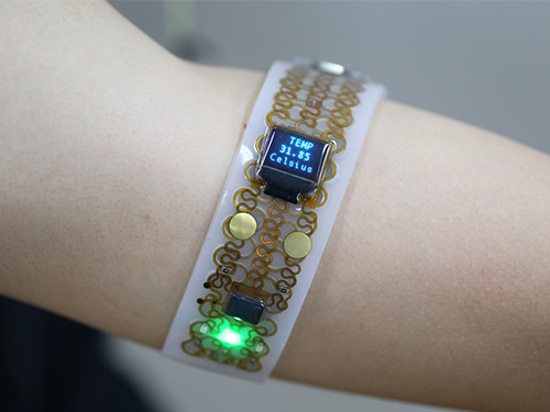 Transformative Electronics Systems to Broaden Wearable Applications
Imagine a handheld electronic gadget that can soften and deform when attached to our skin. This will be the future of electronics we all dreamed of. A research team at KAIST says their new platform called 'Transformative Electronics Systems' will open a new class of electronics, allowing reconfigurable electronic interfaces to be optimized for a variety of applications.
A team working under Professor Jae-Woong Jeong from the School of Electrical Engineering at KAIST has invented a multifunctional electronic platform that can mechanically transform its shape, flexibility, and stretchability. This platform, which was reported in Science Advances, allows users to seamlessly and precisely tune its stiffness and shape.
"This new class of electronics will not only offer robust, convenient interfaces for use in both tabletop or handheld setups, but also allow seamless integration with the skin when applied onto our bodies," said Professor Jeong.
The transformative electronics consist of a special gallium metal structure, hermetically encapsulated and sealed within a soft silicone material, combined with electronics that are designed to be flexible and stretchable. The mechanical transformation of the electronic systems is specifically triggered by temperature change events controlled by the user.
"Gallium is an interesting key material. It is biocompatible, has high rigidity in solid form, and melts at a temperature comparable to the skin's temperature," said lead author Sang-Hyuk Byun, a researcher at KAIST.
Once the transformative electronic platform comes in contact with a human body, the gallium metal encapsulated inside the silicone changes to a liquid state and softens the whole electronic structure, making it stretchable, flexible, and wearable. The gallium metal then solidifies again once the structure is peeled off the skin, making the electronic circuits stiff and stable. When flexible electronic circuits were integrated onto these transformative platforms, it empowered them with the ability to become either flexible and stretchable or rigid.
"This technology could not have been achieved without interdisciplinary efforts," said co-lead author Joo Yong Sim, who is a researcher with ETRI. "We worked together with electrical, mechanical, and biomedical engineers, as well as material scientists and neuroscientists to make this breakthrough."
This universal electronics platform allowed researchers to demonstrate applications that were highly adaptable and customizable, such as a multi-purpose personal electronics with variable stiffness and stretchability, a pressure sensor with tuneable bandwidth and sensitivity, and a neural probe that softens upon implantation into brain tissue.
Applicable for both traditional and emerging electronics technologies, this breakthrough can potentially reshape the consumer electronics industry, especially in the biomedical and robotic domains. The researchers believe that with further development, this novel electronics technology can significantly impact the way we use electronics in our daily life.
< Transformative electronics in soft mode,which becomes wearable for outdoor applications.>
Video Material:
https://youtu.be/im0J18TfShk
Publication: Sang-Hyuk Byun, Joo Yong Sim, Zhanan Zhou, Juhyun Lee, Raza Qazi, Marie C. Walicki, Kyle E. Parker, Matthew P. Haney, Su Hwan Choi, Ahnsei Shon, Graydon B. Gereau, John Bilbily, Shuo Li, Yuhao Liu, Woon-Hong Yeo, Jordan G. McCall, Jianliang Xiao, and Jae-Woong Jeong. 2019. Mechanically transformative electronics, sensors, and implantable devices. Science Advances. Volume 5. No. 11. 12 pages. https://doi.org/10.1126/sciadv.aay0418
Link to download the full-text paper: https://advances.sciencemag.org/content/advances/5/11/eaay0418.full.pdf
Profile: Prof. Jae-Woong Jeong, PhD jjeong1@kaist.ac.kr
https://www.jeongresearch.org/
Professor
Bio-Integrated Electronics and Systems Laboratory
School of Electrical Engineering
Korea Advanced Institute of Science and Technology (KAIST) https://www.kaist.ac.kr
Daejeon 34141, Korea
Profile: Sang-Hyuk Byun, PhD Candidate
shbun95@kaist.ac.kr
(END)
2020.01.31 View 11182
Transformative Electronics Systems to Broaden Wearable Applications
Imagine a handheld electronic gadget that can soften and deform when attached to our skin. This will be the future of electronics we all dreamed of. A research team at KAIST says their new platform called 'Transformative Electronics Systems' will open a new class of electronics, allowing reconfigurable electronic interfaces to be optimized for a variety of applications.
A team working under Professor Jae-Woong Jeong from the School of Electrical Engineering at KAIST has invented a multifunctional electronic platform that can mechanically transform its shape, flexibility, and stretchability. This platform, which was reported in Science Advances, allows users to seamlessly and precisely tune its stiffness and shape.
"This new class of electronics will not only offer robust, convenient interfaces for use in both tabletop or handheld setups, but also allow seamless integration with the skin when applied onto our bodies," said Professor Jeong.
The transformative electronics consist of a special gallium metal structure, hermetically encapsulated and sealed within a soft silicone material, combined with electronics that are designed to be flexible and stretchable. The mechanical transformation of the electronic systems is specifically triggered by temperature change events controlled by the user.
"Gallium is an interesting key material. It is biocompatible, has high rigidity in solid form, and melts at a temperature comparable to the skin's temperature," said lead author Sang-Hyuk Byun, a researcher at KAIST.
Once the transformative electronic platform comes in contact with a human body, the gallium metal encapsulated inside the silicone changes to a liquid state and softens the whole electronic structure, making it stretchable, flexible, and wearable. The gallium metal then solidifies again once the structure is peeled off the skin, making the electronic circuits stiff and stable. When flexible electronic circuits were integrated onto these transformative platforms, it empowered them with the ability to become either flexible and stretchable or rigid.
"This technology could not have been achieved without interdisciplinary efforts," said co-lead author Joo Yong Sim, who is a researcher with ETRI. "We worked together with electrical, mechanical, and biomedical engineers, as well as material scientists and neuroscientists to make this breakthrough."
This universal electronics platform allowed researchers to demonstrate applications that were highly adaptable and customizable, such as a multi-purpose personal electronics with variable stiffness and stretchability, a pressure sensor with tuneable bandwidth and sensitivity, and a neural probe that softens upon implantation into brain tissue.
Applicable for both traditional and emerging electronics technologies, this breakthrough can potentially reshape the consumer electronics industry, especially in the biomedical and robotic domains. The researchers believe that with further development, this novel electronics technology can significantly impact the way we use electronics in our daily life.
< Transformative electronics in soft mode,which becomes wearable for outdoor applications.>
Video Material:
https://youtu.be/im0J18TfShk
Publication: Sang-Hyuk Byun, Joo Yong Sim, Zhanan Zhou, Juhyun Lee, Raza Qazi, Marie C. Walicki, Kyle E. Parker, Matthew P. Haney, Su Hwan Choi, Ahnsei Shon, Graydon B. Gereau, John Bilbily, Shuo Li, Yuhao Liu, Woon-Hong Yeo, Jordan G. McCall, Jianliang Xiao, and Jae-Woong Jeong. 2019. Mechanically transformative electronics, sensors, and implantable devices. Science Advances. Volume 5. No. 11. 12 pages. https://doi.org/10.1126/sciadv.aay0418
Link to download the full-text paper: https://advances.sciencemag.org/content/advances/5/11/eaay0418.full.pdf
Profile: Prof. Jae-Woong Jeong, PhD jjeong1@kaist.ac.kr
https://www.jeongresearch.org/
Professor
Bio-Integrated Electronics and Systems Laboratory
School of Electrical Engineering
Korea Advanced Institute of Science and Technology (KAIST) https://www.kaist.ac.kr
Daejeon 34141, Korea
Profile: Sang-Hyuk Byun, PhD Candidate
shbun95@kaist.ac.kr
(END)
2020.01.31 View 11182 -
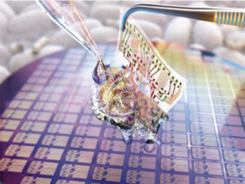 Wafer-Scale Multilayer Fabrication of Silk Fibroin-Based Microelectronics
A KAIST research team developed a novel fabrication method for the multilayer processing of silk-based microelectronics. This technology for creating a biodegradable silk fibroin film allows microfabrication with polymer or metal structures manufactured from photolithography. It can be a key technology in the implementation of silk fibroin-based biodegradable electronic devices or localized drug delivery through silk fibroin patterns.
Silk fibroins are biocompatible, biodegradable, transparent, and flexible, which makes them excellent candidates for implantable biomedical devices, and they have also been used as biodegradable films and functional microstructures in biomedical applications. However, conventional microfabrication processes require strong etching solutions and solvents to modify the structure of silk fibroins.
To prevent the silk fibroin from being damaged during the process, Professor Hyunjoo J. Lee from the School of Electrical Engineering and her team came up with a novel process, named aluminum hard mask on silk fibroin (AMoS), which is capable of micropatterning multiple layers composed of both fibroin and inorganic materials, such as metal and dielectrics with high-precision microscale alignment. The AMoS process can make silk fibroin patterns on devices, or make patterns on silk fibroin thin films with other materials by using photolithography, which is a core technology in the current microfabrication process.
The team successfully cultured primary neurons on the processed silk fibroin micro-patterns, and confirmed that silk fibroin has excellent biocompatibility before and after the fabrication process and that it also can be applied to implanted biological devices.
Through this technology, the team realized the multilayer micropatterning of fibroin films on a silk fibroin substrate and fabricated a biodegradable microelectric circuit consisting of resistors and silk fibroin dielectric capacitors in a silicon wafer with large areas.
They also used this technology to position the micro-pattern of the silk fibroin thin film closer to the flexible polymer-based brain electrode, and confirmed the dye molecules mounted on the silk fibroin were transferred successfully from the micropatterns.
Professor Lee said, “This technology facilitates wafer-scale, large-area processing of sensitive materials. We expect it to be applied to a wide range of biomedical devices in the future. Using the silk fibroin with micro-patterned brain electrodes can open up many new possibilities in research on brain circuits by mounting drugs that restrict or promote brain cell activities.”
This research, in collaboration with Dr. Nakwon Choi from KIST and led by PhD candidate Geon Kook, was published in ACS AMI (10.1021/acsami.8b13170) on January 16, 2019.
Figure 1. The cover page of ACS AMI
Figure 2. Fibroin microstructures and metal patterns on a fibroin produced by using the AMoS mask.
Figure 3. Biocompatibility assessment of the AMoS Process. Top: Schematics image of a) fibroin-coated silicon b) fibroin-pattered silicon and c) gold-patterned fibroin. Bottom: Representative confocal microscopy images of live (green) and dead (red) primary cortical neurons cultured on the substrates.
2019.03.15 View 26153
Wafer-Scale Multilayer Fabrication of Silk Fibroin-Based Microelectronics
A KAIST research team developed a novel fabrication method for the multilayer processing of silk-based microelectronics. This technology for creating a biodegradable silk fibroin film allows microfabrication with polymer or metal structures manufactured from photolithography. It can be a key technology in the implementation of silk fibroin-based biodegradable electronic devices or localized drug delivery through silk fibroin patterns.
Silk fibroins are biocompatible, biodegradable, transparent, and flexible, which makes them excellent candidates for implantable biomedical devices, and they have also been used as biodegradable films and functional microstructures in biomedical applications. However, conventional microfabrication processes require strong etching solutions and solvents to modify the structure of silk fibroins.
To prevent the silk fibroin from being damaged during the process, Professor Hyunjoo J. Lee from the School of Electrical Engineering and her team came up with a novel process, named aluminum hard mask on silk fibroin (AMoS), which is capable of micropatterning multiple layers composed of both fibroin and inorganic materials, such as metal and dielectrics with high-precision microscale alignment. The AMoS process can make silk fibroin patterns on devices, or make patterns on silk fibroin thin films with other materials by using photolithography, which is a core technology in the current microfabrication process.
The team successfully cultured primary neurons on the processed silk fibroin micro-patterns, and confirmed that silk fibroin has excellent biocompatibility before and after the fabrication process and that it also can be applied to implanted biological devices.
Through this technology, the team realized the multilayer micropatterning of fibroin films on a silk fibroin substrate and fabricated a biodegradable microelectric circuit consisting of resistors and silk fibroin dielectric capacitors in a silicon wafer with large areas.
They also used this technology to position the micro-pattern of the silk fibroin thin film closer to the flexible polymer-based brain electrode, and confirmed the dye molecules mounted on the silk fibroin were transferred successfully from the micropatterns.
Professor Lee said, “This technology facilitates wafer-scale, large-area processing of sensitive materials. We expect it to be applied to a wide range of biomedical devices in the future. Using the silk fibroin with micro-patterned brain electrodes can open up many new possibilities in research on brain circuits by mounting drugs that restrict or promote brain cell activities.”
This research, in collaboration with Dr. Nakwon Choi from KIST and led by PhD candidate Geon Kook, was published in ACS AMI (10.1021/acsami.8b13170) on January 16, 2019.
Figure 1. The cover page of ACS AMI
Figure 2. Fibroin microstructures and metal patterns on a fibroin produced by using the AMoS mask.
Figure 3. Biocompatibility assessment of the AMoS Process. Top: Schematics image of a) fibroin-coated silicon b) fibroin-pattered silicon and c) gold-patterned fibroin. Bottom: Representative confocal microscopy images of live (green) and dead (red) primary cortical neurons cultured on the substrates.
2019.03.15 View 26153