3D+holographic
-
 KAIST builds a high-resolution 3D holographic sensor using a single mask
Holographic cameras can provide more realistic images than ordinary cameras thanks to their ability to acquire 3D information about objects. However, existing holographic cameras use interferometers that measure the wavelength and refraction of light through the interference of light waves, which makes them complex and sensitive to their surrounding environment.
On August 23, a KAIST research team led by Professor YongKeun Park from the Department of Physics announced a new leap forward in 3D holographic imaging sensor technology.
The team proposed an innovative holographic camera technology that does not use complex interferometry. Instead, it uses a mask to precisely measure the phase information of light and reconstruct the 3D information of an object with higher accuracy.
< Figure 1. Structure and principle of the proposed holographic camera. The amplitude and phase information of light scattered from a holographic camera can be measured. >
The team used a mask that fulfills certain mathematical conditions and incorporated it into an ordinary camera, and the light scattered from a laser is measured through the mask and analyzed using a computer. This does not require a complex interferometer and allows the phase information of light to be collected through a simplified optical system. With this technique, the mask that is placed between the two lenses and behind an object plays an important role. The mask selectively filters specific parts of light,, and the intensity of the light passing through the lens can be measured using an ordinary commercial camera. This technique combines the image data received from the camera with the unique pattern received from the mask and reconstructs an object’s precise 3D information using an algorithm.
This method allows a high-resolution 3D image of an object to be captured in any position. In practical situations, one can construct a laser-based holographic 3D image sensor by adding a mask with a simple design to a general image sensor. This makes the design and construction of the optical system much easier. In particular, this novel technology can capture high-resolution holographic images of objects moving at high speeds, which widens its potential field of application.
< Figure 2. A moving doll captured by a conventional camera and the proposed holographic camera. When taking a picture without focusing on the object, only a blurred image of the doll can be obtained from a general camera, but the proposed holographic camera can restore the blurred image of the doll into a clear image. >
The results of this study, conducted by Dr. Jeonghun Oh from the KAIST Department of Physics as the first author, were published in Nature Communications on August 12 under the title, "Non-interferometric stand-alone single-shot holographic camera using reciprocal diffractive imaging".
Dr. Oh said, “The holographic camera module we are suggesting can be built by adding a filter to an ordinary camera, which would allow even non-experts to handle it easily in everyday life if it were to be commercialized.” He added, “In particular, it is a promising candidate with the potential to replace existing remote sensing technologies.”
This research was supported by the National Research Foundation’s Leader Research Project, the Korean Ministry of Science and ICT’s Core Hologram Technology Support Project, and the Nano and Material Technology Development Project.
2023.09.05 View 7004
KAIST builds a high-resolution 3D holographic sensor using a single mask
Holographic cameras can provide more realistic images than ordinary cameras thanks to their ability to acquire 3D information about objects. However, existing holographic cameras use interferometers that measure the wavelength and refraction of light through the interference of light waves, which makes them complex and sensitive to their surrounding environment.
On August 23, a KAIST research team led by Professor YongKeun Park from the Department of Physics announced a new leap forward in 3D holographic imaging sensor technology.
The team proposed an innovative holographic camera technology that does not use complex interferometry. Instead, it uses a mask to precisely measure the phase information of light and reconstruct the 3D information of an object with higher accuracy.
< Figure 1. Structure and principle of the proposed holographic camera. The amplitude and phase information of light scattered from a holographic camera can be measured. >
The team used a mask that fulfills certain mathematical conditions and incorporated it into an ordinary camera, and the light scattered from a laser is measured through the mask and analyzed using a computer. This does not require a complex interferometer and allows the phase information of light to be collected through a simplified optical system. With this technique, the mask that is placed between the two lenses and behind an object plays an important role. The mask selectively filters specific parts of light,, and the intensity of the light passing through the lens can be measured using an ordinary commercial camera. This technique combines the image data received from the camera with the unique pattern received from the mask and reconstructs an object’s precise 3D information using an algorithm.
This method allows a high-resolution 3D image of an object to be captured in any position. In practical situations, one can construct a laser-based holographic 3D image sensor by adding a mask with a simple design to a general image sensor. This makes the design and construction of the optical system much easier. In particular, this novel technology can capture high-resolution holographic images of objects moving at high speeds, which widens its potential field of application.
< Figure 2. A moving doll captured by a conventional camera and the proposed holographic camera. When taking a picture without focusing on the object, only a blurred image of the doll can be obtained from a general camera, but the proposed holographic camera can restore the blurred image of the doll into a clear image. >
The results of this study, conducted by Dr. Jeonghun Oh from the KAIST Department of Physics as the first author, were published in Nature Communications on August 12 under the title, "Non-interferometric stand-alone single-shot holographic camera using reciprocal diffractive imaging".
Dr. Oh said, “The holographic camera module we are suggesting can be built by adding a filter to an ordinary camera, which would allow even non-experts to handle it easily in everyday life if it were to be commercialized.” He added, “In particular, it is a promising candidate with the potential to replace existing remote sensing technologies.”
This research was supported by the National Research Foundation’s Leader Research Project, the Korean Ministry of Science and ICT’s Core Hologram Technology Support Project, and the Nano and Material Technology Development Project.
2023.09.05 View 7004 -
 Label-Free Multiplexed Microtomography of Endogenous Subcellular Dynamics Using Deep Learning
AI-based holographic microscopy allows molecular imaging without introducing exogenous labeling agents
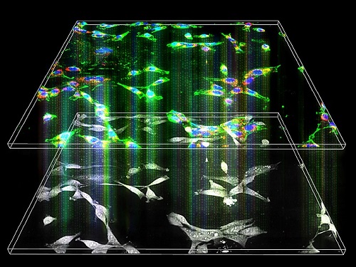
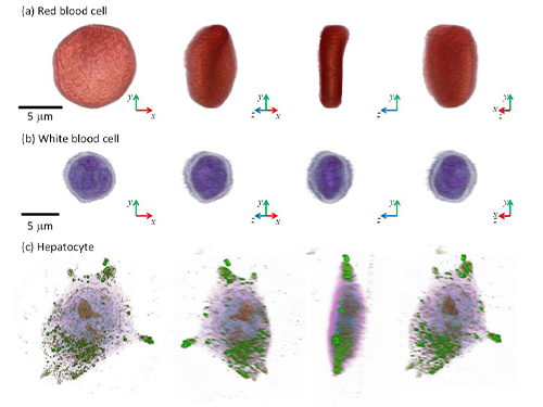
A research team upgraded the 3D microtomography observing dynamics of label-free live cells in multiplexed fluorescence imaging. The AI-powered 3D holotomographic microscopy extracts various molecular information from live unlabeled biological cells in real time without exogenous labeling or staining agents.
Professor YongKeum Park’s team and the startup Tomocube encoded 3D refractive index tomograms using the refractive index as a means of measurement. Then they decoded the information with a deep learning-based model that infers multiple 3D fluorescence tomograms from the refractive index measurements of the corresponding subcellular targets, thereby achieving multiplexed micro tomography. This study was reported in Nature Cell Biology online on December 7, 2021.
Fluorescence microscopy is the most widely used optical microscopy technique due to its high biochemical specificity. However, it needs to genetically manipulate or to stain cells with fluorescent labels in order to express fluorescent proteins. These labeling processes inevitably affect the intrinsic physiology of cells. It also has challenges in long-term measuring due to photobleaching and phototoxicity. The overlapped spectra of multiplexed fluorescence signals also hinder the viewing of various structures at the same time. More critically, it took several hours to observe the cells after preparing them.
3D holographic microscopy, also known as holotomography, is providing new ways to quantitatively image live cells without pretreatments such as staining. Holotomography can accurately and quickly measure the morphological and structural information of cells, but only provides limited biochemical and molecular information.
The 'AI microscope' created in this process takes advantage of the features of both holographic microscopy and fluorescence microscopy. That is, a specific image from a fluorescence microscope can be obtained without a fluorescent label. Therefore, the microscope can observe many types of cellular structures in their natural state in 3D and at the same time as fast as one millisecond, and long-term measurements over several days are also possible.
The Tomocube-KAIST team showed that fluorescence images can be directly and precisely predicted from holotomographic images in various cells and conditions. Using the quantitative relationship between the spatial distribution of the refractive index found by AI and the major structures in cells, it was possible to decipher the spatial distribution of the refractive index. And surprisingly, it confirmed that this relationship is constant regardless of cell type.
Professor Park said, “We were able to develop a new concept microscope that combines the advantages of several microscopes with the multidisciplinary research of AI, optics, and biology. It will be immediately applicable for new types of cells not included in the existing data and is expected to be widely applicable for various biological and medical research.”
When comparing the molecular image information extracted by AI with the molecular image information physically obtained by fluorescence staining in 3D space, it showed a 97% or more conformity, which is a level that is difficult to distinguish with the naked eye.
“Compared to the sub-60% accuracy of the fluorescence information extracted from the model developed by the Google AI team, it showed significantly higher performance,” Professor Park added.
This work was supported by the KAIST Up program, the BK21+ program, Tomocube, the National Research Foundation of Korea, and the Ministry of Science and ICT, and the Ministry of Health & Welfare.
-Publication
Hyun-seok Min, Won-Do Heo, YongKeun Park, et al. “Label-free multiplexed microtomography of endogenous subcellular dynamics using generalizable deep learning,” Nature Cell Biology (doi.org/10.1038/s41556-021-00802-x) published online December 07 2021.
-Profile
Professor YongKeun Park
Biomedical Optics Laboratory
Department of Physics
KAIST
2022.02.09 View 10756
Label-Free Multiplexed Microtomography of Endogenous Subcellular Dynamics Using Deep Learning
AI-based holographic microscopy allows molecular imaging without introducing exogenous labeling agents
A research team upgraded the 3D microtomography observing dynamics of label-free live cells in multiplexed fluorescence imaging. The AI-powered 3D holotomographic microscopy extracts various molecular information from live unlabeled biological cells in real time without exogenous labeling or staining agents.
Professor YongKeum Park’s team and the startup Tomocube encoded 3D refractive index tomograms using the refractive index as a means of measurement. Then they decoded the information with a deep learning-based model that infers multiple 3D fluorescence tomograms from the refractive index measurements of the corresponding subcellular targets, thereby achieving multiplexed micro tomography. This study was reported in Nature Cell Biology online on December 7, 2021.
Fluorescence microscopy is the most widely used optical microscopy technique due to its high biochemical specificity. However, it needs to genetically manipulate or to stain cells with fluorescent labels in order to express fluorescent proteins. These labeling processes inevitably affect the intrinsic physiology of cells. It also has challenges in long-term measuring due to photobleaching and phototoxicity. The overlapped spectra of multiplexed fluorescence signals also hinder the viewing of various structures at the same time. More critically, it took several hours to observe the cells after preparing them.
3D holographic microscopy, also known as holotomography, is providing new ways to quantitatively image live cells without pretreatments such as staining. Holotomography can accurately and quickly measure the morphological and structural information of cells, but only provides limited biochemical and molecular information.
The 'AI microscope' created in this process takes advantage of the features of both holographic microscopy and fluorescence microscopy. That is, a specific image from a fluorescence microscope can be obtained without a fluorescent label. Therefore, the microscope can observe many types of cellular structures in their natural state in 3D and at the same time as fast as one millisecond, and long-term measurements over several days are also possible.
The Tomocube-KAIST team showed that fluorescence images can be directly and precisely predicted from holotomographic images in various cells and conditions. Using the quantitative relationship between the spatial distribution of the refractive index found by AI and the major structures in cells, it was possible to decipher the spatial distribution of the refractive index. And surprisingly, it confirmed that this relationship is constant regardless of cell type.
Professor Park said, “We were able to develop a new concept microscope that combines the advantages of several microscopes with the multidisciplinary research of AI, optics, and biology. It will be immediately applicable for new types of cells not included in the existing data and is expected to be widely applicable for various biological and medical research.”
When comparing the molecular image information extracted by AI with the molecular image information physically obtained by fluorescence staining in 3D space, it showed a 97% or more conformity, which is a level that is difficult to distinguish with the naked eye.
“Compared to the sub-60% accuracy of the fluorescence information extracted from the model developed by the Google AI team, it showed significantly higher performance,” Professor Park added.
This work was supported by the KAIST Up program, the BK21+ program, Tomocube, the National Research Foundation of Korea, and the Ministry of Science and ICT, and the Ministry of Health & Welfare.
-Publication
Hyun-seok Min, Won-Do Heo, YongKeun Park, et al. “Label-free multiplexed microtomography of endogenous subcellular dynamics using generalizable deep learning,” Nature Cell Biology (doi.org/10.1038/s41556-021-00802-x) published online December 07 2021.
-Profile
Professor YongKeun Park
Biomedical Optics Laboratory
Department of Physics
KAIST
2022.02.09 View 10756 -
 3D Visualization and Quantification of Bioplastic PHA in a Living Bacterial Cell
3D holographic microscopy leads to in-depth analysis of bacterial cells accumulating the bacterial bioplastic, polyhydroxyalkanoate (PHA)
A research team at KAIST has observed how bioplastic granule is being accumulated in living bacteria cells through 3D holographic microscopy. Their 3D imaging and quantitative analysis of the bioplastic ‘polyhydroxyalkanoate’ (PHA) via optical diffraction tomography provides insights into biosynthesizing sustainable substitutes for petroleum-based plastics.
The bio-degradable polyester polyhydroxyalkanoate (PHA) is being touted as an eco-friendly bioplastic to replace existing synthetic plastics. While carrying similar properties to general-purpose plastics such as polyethylene and polypropylene, PHA can be used in various industrial applications such as container packaging and disposable products.
PHA is synthesized by numerous bacteria as an energy and carbon storage material under unbalanced growth conditions in the presence of excess carbon sources. PHA exists in the form of insoluble granules in the cytoplasm. Previous studies on investigating in vivo PHA granules have been performed by using fluorescence microscopy, transmission electron microscopy (TEM), and electron cryotomography.
These techniques have generally relied on the statistical analysis of multiple 2D snapshots of fixed cells or the short-time monitoring of the cells. For the TEM analysis, cells need to be fixed and sectioned, and thus the investigation of living cells was not possible. Fluorescence-based techniques require fluorescence labeling or dye staining. Thus, indirect imaging with the use of reporter proteins cannot show the native state of PHAs or cells, and invasive exogenous dyes can affect the physiology and viability of the cells. Therefore, it was difficult to fully understand the formation of PHA granules in cells due to the technical limitations, and thus several mechanism models based on the observations have been only proposed.
The team of metabolic engineering researchers led by Distinguished Professor Sang Yup Lee and Physics Professor YongKeun Park, who established the startup Tomocube with his 3D holographic microscopy, reported the results of 3D quantitative label-free analysis of PHA granules in individual live bacterial cells by measuring the refractive index distributions using optical diffraction tomography. The formation and growth of PHA granules in the cells of Cupriavidus necator, the most-studied native PHA (specifically, poly(3-hydroxybutyrate), also known as PHB) producer, and recombinant Escherichia coli harboring C. necator PHB biosynthesis pathway were comparatively examined.
From the reconstructed 3D refractive index distribution of the cells, the team succeeded in the 3D visualization and quantitative analysis of cells and intracellular PHA granules at a single-cell level. In particular, the team newly presented the concept of “in vivo PHA granule density.” Through the statistical analysis of hundreds of single cells accumulating PHA granules, the distinctive differences of density and localization of PHA granules in the two micro-organisms were found. Furthermore, the team identified the key protein that plays a major role in making the difference that enabled the characteristics of PHA granules in the recombinant E. coli to become similar to those of C. necator.
The research team also presented 3D time-lapse movies showing the actual processes of PHA granule formation combined with cell growth and division. Movies showing the living cells synthesizing and accumulating PHA granules in their native state had never been reported before.
Professor Lee said, “This study provides insights into the morphological and physical characteristics of in vivo PHA as well as the unique mechanisms of PHA granule formation that undergo the phase transition from soluble monomers into the insoluble polymer, followed by granule formation. Through this study, a deeper understanding of PHA granule formation within the bacterial cells is now possible, which has great significance in that a convergence study of biology and physics was achieved. This study will help develop various bioplastics production processes in the future.”
This work was supported by the Technology Development Program to Solve Climate Changes on Systems Metabolic Engineering for Biorefineries (Grants NRF-2012M1A2A2026556 and NRF-2012M1A2A2026557) and the Bio & Medical Technology Development Program (Grant No. 2021M3A9I4022740) from the Ministry of Science and ICT (MSIT) through the National Research Foundation (NRF) of Korea to S.Y.L. This work was also supported by the KAIST Cross-Generation Collaborative Laboratory project.
-PublicationSo Young Choi, Jeonghun Oh, JaeHwang Jung, YongKeun Park, and Sang Yup Lee. Three-dimensional label-free visualization and quantification of polyhydroxyalkanoates in individualbacterial cell in its native state. PNAS(https://doi.org./10.1073/pnas.2103956118)
-ProfileDistinguished Professor Sang Yup LeeMetabolic Engineering and Synthetic Biologyhttp://mbel.kaist.ac.kr/
Department of Chemical and Biomolecular Engineering KAIST
Endowed Chair Professor YongKeun ParkBiomedical Optics Laboratoryhttps://bmokaist.wordpress.com/
Department of PhysicsKAIST
2021.07.28 View 13266
3D Visualization and Quantification of Bioplastic PHA in a Living Bacterial Cell
3D holographic microscopy leads to in-depth analysis of bacterial cells accumulating the bacterial bioplastic, polyhydroxyalkanoate (PHA)
A research team at KAIST has observed how bioplastic granule is being accumulated in living bacteria cells through 3D holographic microscopy. Their 3D imaging and quantitative analysis of the bioplastic ‘polyhydroxyalkanoate’ (PHA) via optical diffraction tomography provides insights into biosynthesizing sustainable substitutes for petroleum-based plastics.
The bio-degradable polyester polyhydroxyalkanoate (PHA) is being touted as an eco-friendly bioplastic to replace existing synthetic plastics. While carrying similar properties to general-purpose plastics such as polyethylene and polypropylene, PHA can be used in various industrial applications such as container packaging and disposable products.
PHA is synthesized by numerous bacteria as an energy and carbon storage material under unbalanced growth conditions in the presence of excess carbon sources. PHA exists in the form of insoluble granules in the cytoplasm. Previous studies on investigating in vivo PHA granules have been performed by using fluorescence microscopy, transmission electron microscopy (TEM), and electron cryotomography.
These techniques have generally relied on the statistical analysis of multiple 2D snapshots of fixed cells or the short-time monitoring of the cells. For the TEM analysis, cells need to be fixed and sectioned, and thus the investigation of living cells was not possible. Fluorescence-based techniques require fluorescence labeling or dye staining. Thus, indirect imaging with the use of reporter proteins cannot show the native state of PHAs or cells, and invasive exogenous dyes can affect the physiology and viability of the cells. Therefore, it was difficult to fully understand the formation of PHA granules in cells due to the technical limitations, and thus several mechanism models based on the observations have been only proposed.
The team of metabolic engineering researchers led by Distinguished Professor Sang Yup Lee and Physics Professor YongKeun Park, who established the startup Tomocube with his 3D holographic microscopy, reported the results of 3D quantitative label-free analysis of PHA granules in individual live bacterial cells by measuring the refractive index distributions using optical diffraction tomography. The formation and growth of PHA granules in the cells of Cupriavidus necator, the most-studied native PHA (specifically, poly(3-hydroxybutyrate), also known as PHB) producer, and recombinant Escherichia coli harboring C. necator PHB biosynthesis pathway were comparatively examined.
From the reconstructed 3D refractive index distribution of the cells, the team succeeded in the 3D visualization and quantitative analysis of cells and intracellular PHA granules at a single-cell level. In particular, the team newly presented the concept of “in vivo PHA granule density.” Through the statistical analysis of hundreds of single cells accumulating PHA granules, the distinctive differences of density and localization of PHA granules in the two micro-organisms were found. Furthermore, the team identified the key protein that plays a major role in making the difference that enabled the characteristics of PHA granules in the recombinant E. coli to become similar to those of C. necator.
The research team also presented 3D time-lapse movies showing the actual processes of PHA granule formation combined with cell growth and division. Movies showing the living cells synthesizing and accumulating PHA granules in their native state had never been reported before.
Professor Lee said, “This study provides insights into the morphological and physical characteristics of in vivo PHA as well as the unique mechanisms of PHA granule formation that undergo the phase transition from soluble monomers into the insoluble polymer, followed by granule formation. Through this study, a deeper understanding of PHA granule formation within the bacterial cells is now possible, which has great significance in that a convergence study of biology and physics was achieved. This study will help develop various bioplastics production processes in the future.”
This work was supported by the Technology Development Program to Solve Climate Changes on Systems Metabolic Engineering for Biorefineries (Grants NRF-2012M1A2A2026556 and NRF-2012M1A2A2026557) and the Bio & Medical Technology Development Program (Grant No. 2021M3A9I4022740) from the Ministry of Science and ICT (MSIT) through the National Research Foundation (NRF) of Korea to S.Y.L. This work was also supported by the KAIST Cross-Generation Collaborative Laboratory project.
-PublicationSo Young Choi, Jeonghun Oh, JaeHwang Jung, YongKeun Park, and Sang Yup Lee. Three-dimensional label-free visualization and quantification of polyhydroxyalkanoates in individualbacterial cell in its native state. PNAS(https://doi.org./10.1073/pnas.2103956118)
-ProfileDistinguished Professor Sang Yup LeeMetabolic Engineering and Synthetic Biologyhttp://mbel.kaist.ac.kr/
Department of Chemical and Biomolecular Engineering KAIST
Endowed Chair Professor YongKeun ParkBiomedical Optics Laboratoryhttps://bmokaist.wordpress.com/
Department of PhysicsKAIST
2021.07.28 View 13266 -
 Deep-Learning and 3D Holographic Microscopy Beats Scientists at Analyzing Cancer Immunotherapy
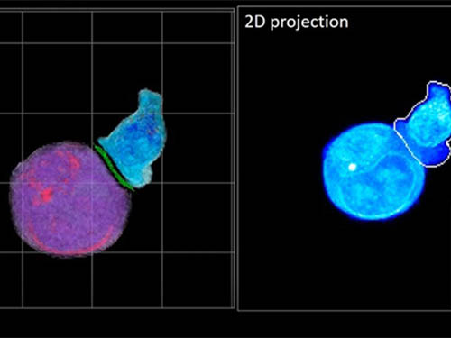
Live tracking and analyzing of the dynamics of chimeric antigen receptor (CAR) T-cells targeting cancer cells can open new avenues for the development of cancer immunotherapy. However, imaging via conventional microscopy approaches can result in cellular damage, and assessments of cell-to-cell interactions are extremely difficult and labor-intensive. When researchers applied deep learning and 3D holographic microscopy to the task, however, they not only avoided these difficultues but found that AI was better at it than humans were.
Artificial intelligence (AI) is helping researchers decipher images from a new holographic microscopy technique needed to investigate a key process in cancer immunotherapy “live” as it takes place. The AI transformed work that, if performed manually by scientists, would otherwise be incredibly labor-intensive and time-consuming into one that is not only effortless but done better than they could have done it themselves. The research, conducted by the team of Professor YongKeun Park from the Department of Physics, appeared in the journal eLife last December.
A critical stage in the development of the human immune system’s ability to respond not just generally to any invader (such as pathogens or cancer cells) but specifically to that particular type of invader and remember it should it attempt to invade again is the formation of a junction between an immune cell called a T-cell and a cell that presents the antigen, or part of the invader that is causing the problem, to it. This process is like when a picture of a suspect is sent to a police car so that the officers can recognize the criminal they are trying to track down. The junction between the two cells, called the immunological synapse, or IS, is the key process in teaching the immune system how to recognize a specific type of invader.
Since the formation of the IS junction is such a critical step for the initiation of an antigen-specific immune response, various techniques allowing researchers to observe the process as it happens have been used to study its dynamics. Most of these live imaging techniques rely on fluorescence microscopy, where genetic tweaking causes part of a protein from a cell to fluoresce, in turn allowing the subject to be tracked via fluorescence rather than via the reflected light used in many conventional microscopy techniques.
However, fluorescence-based imaging can suffer from effects such as photo-bleaching and photo-toxicity, preventing the assessment of dynamic changes in the IS junction process over the long term. Fluorescence-based imaging still involves illumination, whereupon the fluorophores (chemical compounds that cause the fluorescence) emit light of a different color. Photo-bleaching or photo-toxicity occur when the subject is exposed to too much illumination, resulting in chemical alteration or cellular damage.
One recent option that does away with fluorescent labelling and thereby avoids such problems is 3D holographic microscopy or holotomography (HT). In this technique, the refractive index (the way that light changes direction when encountering a substance with a different density—why a straw looks like it bends in a glass of water) is recorded in 3D as a hologram.
Until now, HT has been used to study single cells, but never cell-cell interactions involved in immune responses. One of the main reasons is the difficulty of “segmentation,” or distinguishing the different parts of a cell and thus distinguishing between the interacting cells; in other words, deciphering which part belongs to which cell.
Manual segmentation, or marking out the different parts manually, is one option, but it is difficult and time-consuming, especially in three dimensions. To overcome this problem, automatic segmentation has been developed in which simple computer algorithms perform the identification.
“But these basic algorithms often make mistakes,” explained Professor YongKeun Park, “particularly with respect to adjoining segmentation, which of course is exactly what is occurring here in the immune response we’re most interested in.”
So, the researchers applied a deep learning framework to the HT segmentation problem. Deep learning is a type of machine learning in which artificial neural networks based on the human brain recognize patterns in a way that is similar to how humans do this. Regular machine learning requires data as an input that has already been labelled. The AI “learns” by understanding the labeled data and then recognizes the concept that has been labelled when it is fed novel data. For example, AI trained on a thousand images of cats labelled “cat” should be able to recognize a cat the next time it encounters an image with a cat in it. Deep learning involves multiple layers of artificial neural networks attacking much larger, but unlabeled datasets, in which the AI develops its own ‘labels’ for concepts it encounters.
In essence, the deep learning framework that KAIST researchers developed, called DeepIS, came up with its own concepts by which it distinguishes the different parts of the IS junction process. To validate this method, the research team applied it to the dynamics of a particular IS junction formed between chimeric antigen receptor (CAR) T-cells and target cancer cells. They then compared the results to what they would normally have done: the laborious process of performing the segmentation manually. They found not only that DeepIS was able to define areas within the IS with high accuracy, but that the technique was even able to capture information about the total distribution of proteins within the IS that may not have been easily measured using conventional techniques.
“In addition to allowing us to avoid the drudgery of manual segmentation and the problems of photo-bleaching and photo-toxicity, we found that the AI actually did a better job,” Professor Park added.
The next step will be to combine the technique with methods of measuring how much physical force is applied by different parts of the IS junction, such as holographic optical tweezers or traction force microscopy.
-Profile
Professor YongKeun Park
Department of Physics
Biomedical Optics Laboratory
http://bmol.kaist.ac.kr
KAIST
2021.02.24 View 12542
Deep-Learning and 3D Holographic Microscopy Beats Scientists at Analyzing Cancer Immunotherapy
Live tracking and analyzing of the dynamics of chimeric antigen receptor (CAR) T-cells targeting cancer cells can open new avenues for the development of cancer immunotherapy. However, imaging via conventional microscopy approaches can result in cellular damage, and assessments of cell-to-cell interactions are extremely difficult and labor-intensive. When researchers applied deep learning and 3D holographic microscopy to the task, however, they not only avoided these difficultues but found that AI was better at it than humans were.
Artificial intelligence (AI) is helping researchers decipher images from a new holographic microscopy technique needed to investigate a key process in cancer immunotherapy “live” as it takes place. The AI transformed work that, if performed manually by scientists, would otherwise be incredibly labor-intensive and time-consuming into one that is not only effortless but done better than they could have done it themselves. The research, conducted by the team of Professor YongKeun Park from the Department of Physics, appeared in the journal eLife last December.
A critical stage in the development of the human immune system’s ability to respond not just generally to any invader (such as pathogens or cancer cells) but specifically to that particular type of invader and remember it should it attempt to invade again is the formation of a junction between an immune cell called a T-cell and a cell that presents the antigen, or part of the invader that is causing the problem, to it. This process is like when a picture of a suspect is sent to a police car so that the officers can recognize the criminal they are trying to track down. The junction between the two cells, called the immunological synapse, or IS, is the key process in teaching the immune system how to recognize a specific type of invader.
Since the formation of the IS junction is such a critical step for the initiation of an antigen-specific immune response, various techniques allowing researchers to observe the process as it happens have been used to study its dynamics. Most of these live imaging techniques rely on fluorescence microscopy, where genetic tweaking causes part of a protein from a cell to fluoresce, in turn allowing the subject to be tracked via fluorescence rather than via the reflected light used in many conventional microscopy techniques.
However, fluorescence-based imaging can suffer from effects such as photo-bleaching and photo-toxicity, preventing the assessment of dynamic changes in the IS junction process over the long term. Fluorescence-based imaging still involves illumination, whereupon the fluorophores (chemical compounds that cause the fluorescence) emit light of a different color. Photo-bleaching or photo-toxicity occur when the subject is exposed to too much illumination, resulting in chemical alteration or cellular damage.
One recent option that does away with fluorescent labelling and thereby avoids such problems is 3D holographic microscopy or holotomography (HT). In this technique, the refractive index (the way that light changes direction when encountering a substance with a different density—why a straw looks like it bends in a glass of water) is recorded in 3D as a hologram.
Until now, HT has been used to study single cells, but never cell-cell interactions involved in immune responses. One of the main reasons is the difficulty of “segmentation,” or distinguishing the different parts of a cell and thus distinguishing between the interacting cells; in other words, deciphering which part belongs to which cell.
Manual segmentation, or marking out the different parts manually, is one option, but it is difficult and time-consuming, especially in three dimensions. To overcome this problem, automatic segmentation has been developed in which simple computer algorithms perform the identification.
“But these basic algorithms often make mistakes,” explained Professor YongKeun Park, “particularly with respect to adjoining segmentation, which of course is exactly what is occurring here in the immune response we’re most interested in.”
So, the researchers applied a deep learning framework to the HT segmentation problem. Deep learning is a type of machine learning in which artificial neural networks based on the human brain recognize patterns in a way that is similar to how humans do this. Regular machine learning requires data as an input that has already been labelled. The AI “learns” by understanding the labeled data and then recognizes the concept that has been labelled when it is fed novel data. For example, AI trained on a thousand images of cats labelled “cat” should be able to recognize a cat the next time it encounters an image with a cat in it. Deep learning involves multiple layers of artificial neural networks attacking much larger, but unlabeled datasets, in which the AI develops its own ‘labels’ for concepts it encounters.
In essence, the deep learning framework that KAIST researchers developed, called DeepIS, came up with its own concepts by which it distinguishes the different parts of the IS junction process. To validate this method, the research team applied it to the dynamics of a particular IS junction formed between chimeric antigen receptor (CAR) T-cells and target cancer cells. They then compared the results to what they would normally have done: the laborious process of performing the segmentation manually. They found not only that DeepIS was able to define areas within the IS with high accuracy, but that the technique was even able to capture information about the total distribution of proteins within the IS that may not have been easily measured using conventional techniques.
“In addition to allowing us to avoid the drudgery of manual segmentation and the problems of photo-bleaching and photo-toxicity, we found that the AI actually did a better job,” Professor Park added.
The next step will be to combine the technique with methods of measuring how much physical force is applied by different parts of the IS junction, such as holographic optical tweezers or traction force microscopy.
-Profile
Professor YongKeun Park
Department of Physics
Biomedical Optics Laboratory
http://bmol.kaist.ac.kr
KAIST
2021.02.24 View 12542 -
 A Hole in One for Holographic Display
(Professor YongKeun Park)
Researchers have designed an ultrathin display that can project dynamic, multi-coloured, 3D holographic images, according to a study published in Nature Communications.
The system’s critical component is a thin film of titanium filled with tiny holes that precisely correspond with each pixel in a liquid crystal display (LCD) panel. This film acts as a ‘photon sieve’ – each pinhole diffracts light emerging from them widely, resulting in a high-definition 3D image observable from a wide angle.
The entire system is very small: they used a 1.8-inch off-the-shelf LCD panel with a resolution of 1024 x 768. The titanium film, attached to the back of the panel, is a mere 300 nanometres thick.
“Our approach suggests that holographic displays could be projected from thin devices, like a cell phone,” says Professor YongKeun Park, a physicist at KAIST who led the research. The team demonstrated their approach by producing a hologram of a moving, tri-coloured cube.
Specifically, the images are made by pointing differently coloured laser beams made of parallel light rays at the small LCD panel. The photon sieve has a hole for each pixel in the LCD panel. The holes are precisely positioned to correspond to the pixel’s active area. The pinholes diffract the light emerging from them, producing 3D images.
Previous studies from Professor Park’s group have used optical diffusors for the same purpose, but the size of the device was bulky and difficult to be operated, and it took a long period of time to calibrate. In the present work, on the other hand, the group tailored their photon sieve to demonstrate a simple, compact and scalable method for 3D holographic display. This technique can be readily applied to existing LCD displays.
Applications for holograms have been limited by cumbersome techniques, high computation requirements, and poor image quality. Improving current techniques could lead to a wide variety of applications, including 3D cinema viewing without the need for glasses, watching holographic videos on television and smart phone screens.
Figure 1. The actual 3D holographic display, and an electron microscope image of the non-periodic pinholes.
Figure 2. Three-dimensional dynamic color hologram operating at 60 Hz
2019.04.18 View 33013
A Hole in One for Holographic Display
(Professor YongKeun Park)
Researchers have designed an ultrathin display that can project dynamic, multi-coloured, 3D holographic images, according to a study published in Nature Communications.
The system’s critical component is a thin film of titanium filled with tiny holes that precisely correspond with each pixel in a liquid crystal display (LCD) panel. This film acts as a ‘photon sieve’ – each pinhole diffracts light emerging from them widely, resulting in a high-definition 3D image observable from a wide angle.
The entire system is very small: they used a 1.8-inch off-the-shelf LCD panel with a resolution of 1024 x 768. The titanium film, attached to the back of the panel, is a mere 300 nanometres thick.
“Our approach suggests that holographic displays could be projected from thin devices, like a cell phone,” says Professor YongKeun Park, a physicist at KAIST who led the research. The team demonstrated their approach by producing a hologram of a moving, tri-coloured cube.
Specifically, the images are made by pointing differently coloured laser beams made of parallel light rays at the small LCD panel. The photon sieve has a hole for each pixel in the LCD panel. The holes are precisely positioned to correspond to the pixel’s active area. The pinholes diffract the light emerging from them, producing 3D images.
Previous studies from Professor Park’s group have used optical diffusors for the same purpose, but the size of the device was bulky and difficult to be operated, and it took a long period of time to calibrate. In the present work, on the other hand, the group tailored their photon sieve to demonstrate a simple, compact and scalable method for 3D holographic display. This technique can be readily applied to existing LCD displays.
Applications for holograms have been limited by cumbersome techniques, high computation requirements, and poor image quality. Improving current techniques could lead to a wide variety of applications, including 3D cinema viewing without the need for glasses, watching holographic videos on television and smart phone screens.
Figure 1. The actual 3D holographic display, and an electron microscope image of the non-periodic pinholes.
Figure 2. Three-dimensional dynamic color hologram operating at 60 Hz
2019.04.18 View 33013 -
 Professor YongKeun Park Wins the 2018 Fumio Okano Award
(Professor Park)
Professor YongKeun Park from the Department of Physics won the 2018 Fumio Okano Award in recognition of his contributions to 3D display technology development during the annual conference of the International Society for Optics and Photonics (SPIE) held last month in Orlando, Florida in the US.
The Fumio Okano Best 3D Paper Prize is presented annually in memory of Dr. Fumio Okano, a pioneer and innovator of 3D displays who passed away in 2013, for his contributions to the field of 3D TVs and displays. The award is sponsored by NHK-ES.
Professor Park and his team are developing novel technology for measuring and visualizing 3D images by applying random light scattering. He has published numerous papers on 3D holographic camera technology and 3000x enhanced performance of 3D holographic displays in renowned international journals such as Nature Photonics, Nature Communications, and Science Advances. His technology has drawn international attention from renowned media outlets including Newsweek and Forbes.
He has established two startups to commercialize his technology. Tomocube specializes in 3D imaging microscopes using holotomographic technology and the company exports their products to several countries including the US and Japan. The.Wave.Talk is exploring technology for examining pre-existing bacteria anywhere and anytime.
Professor Park’s innovations have already been recognized in and out of KAIST. In February, he was selected as the KAISTian of the Year for his outstanding research, commercialization, and startups. He was also decorated with the National Science Award in April by the Ministry of Science and ICT and the Hong Jin-Ki Innovation Award later in May by the Yumin Cultural Foundation.
Professor Park said, “3D holography is emerging as a significant technology with growing potential and positive impacts on our daily lives. However, the current technology lags far behind the levels displayed in SF movies. We will do our utmost to reach this level with more commercialization."
2018.05.31 View 11097
Professor YongKeun Park Wins the 2018 Fumio Okano Award
(Professor Park)
Professor YongKeun Park from the Department of Physics won the 2018 Fumio Okano Award in recognition of his contributions to 3D display technology development during the annual conference of the International Society for Optics and Photonics (SPIE) held last month in Orlando, Florida in the US.
The Fumio Okano Best 3D Paper Prize is presented annually in memory of Dr. Fumio Okano, a pioneer and innovator of 3D displays who passed away in 2013, for his contributions to the field of 3D TVs and displays. The award is sponsored by NHK-ES.
Professor Park and his team are developing novel technology for measuring and visualizing 3D images by applying random light scattering. He has published numerous papers on 3D holographic camera technology and 3000x enhanced performance of 3D holographic displays in renowned international journals such as Nature Photonics, Nature Communications, and Science Advances. His technology has drawn international attention from renowned media outlets including Newsweek and Forbes.
He has established two startups to commercialize his technology. Tomocube specializes in 3D imaging microscopes using holotomographic technology and the company exports their products to several countries including the US and Japan. The.Wave.Talk is exploring technology for examining pre-existing bacteria anywhere and anytime.
Professor Park’s innovations have already been recognized in and out of KAIST. In February, he was selected as the KAISTian of the Year for his outstanding research, commercialization, and startups. He was also decorated with the National Science Award in April by the Ministry of Science and ICT and the Hong Jin-Ki Innovation Award later in May by the Yumin Cultural Foundation.
Professor Park said, “3D holography is emerging as a significant technology with growing potential and positive impacts on our daily lives. However, the current technology lags far behind the levels displayed in SF movies. We will do our utmost to reach this level with more commercialization."
2018.05.31 View 11097 -
 2017 KAIST Research Day Honors Professor Hoon Sohn
The 2017 KAIST Research Day recognized Professor Hoon Sohn of the Department of Civil and Environmental Engineering as Research Grand Prize Awardee in addition to the 10 most distinguished research achievements of the past year.
The Research Grand Prize recognizes the professor whose comprehensive research performance evaluation indicator is the highest over the past five years. The indicator combines the factors of the number of research contracts, IPR, royalty income, as well as research overhead cost inclusion.
During the ceremony, which was held on May 23, Professor Jun-Ho Oh of the Department of Mechanical Engineering and Professor Sang Yup Lee of the Department of Chemical and Biomolecular Engineering also won the Best Research Award. The two professors had the best scores when evaluating their research performance for one-year periods.
Meanwhile, the Research Innovation Award went to Professor YongKeun Park of the Department of Physics. The Research Innovation Award scores the factors of foreign patent registration, contracts of technological transfer and income from technology fees, technology consultations, and startups and selected Professor Park as the top winner.
Professors Yong Hee Lee of the Department of Physics and Jonghwa Shin of the Department of Material Science won the Convergence Research Award. The Convergence Research Award recognizes the most outstanding research team who created innovative research results for a year.
After the ceremony, President Chen Shiyi of the Southern University of Science and Technology gave a distinguished lecture on the “Global & Entrepreneurial Universities for the Age of the Fourth Industrial Revolution.” the Research Day ceremony, KAIST also presented the ten most distinguished research achievements made by KAIST professors during the last year as follows (Click):
▲ Commercialization of 3D Holographic Microscopy by Professor YongKeun Park of the Department of Physics
▲ Designer Proteins with Chemical Modifications by Professor Hee-Sung Park of the Department of Chemistry
▲ Lanthanum-Catalyzed Synthesis of Microporous 3D Graphene-Like Carbons in a Zeolite Template by Professor Ryong Ryoo of the Department of Chemistry
▲ Complete Prevention of Blood Loss by Self-Sealing Hemostatic Needles by Professor Haeshin Lee of the Department of Chemistry
▲ An Immunological Mechanism for the Contribution of Commensal Microbiota Against Herpes Simplex Virus Infection in Genital Mucosa by Heung Kyu Lee of the Graduate School of Medical Science and Engineering
▲ Development of a Pulse-Echo Laser Ultrasonic Propagation Imaging System by Professor Jung-Ryul Lee of the Department of Aerospace Engineering
▲ Bi-refractive Stereo Imaging for Single-Shot Depth Acquisition by Professor Min H. Kim of the School of Computing
▲ Development of Environment Friendly Geotechnical Construction Material Using Biopolymer by Professor Gye-Chun Cho of the Department of Civil and Environmental Engineering
▲ Protein Delivery Via Engineered Exosomes by Professor Chulhee Choi of the Department of Bio and Brain Engineering
▲ Hot Electron Detection Under Catalytic Reactions by Professor Jeong Young Park of the Graduate School of EEWS.
After the ceremony, President Chen Shiyi of the Southern University of Science and Technology gave a distinguished lecture on the “Global & Entrepreneurial Universities for the Age of the Fourth Industrial Revolution.”
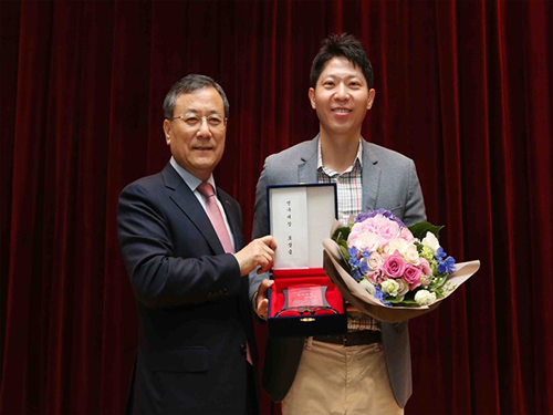
(Photo:President Shin poses with the 2017 KAIST Research Grand Prize Winner Professor Hoon Sohn on May 23.)
2017.05.23 View 13946
2017 KAIST Research Day Honors Professor Hoon Sohn
The 2017 KAIST Research Day recognized Professor Hoon Sohn of the Department of Civil and Environmental Engineering as Research Grand Prize Awardee in addition to the 10 most distinguished research achievements of the past year.
The Research Grand Prize recognizes the professor whose comprehensive research performance evaluation indicator is the highest over the past five years. The indicator combines the factors of the number of research contracts, IPR, royalty income, as well as research overhead cost inclusion.
During the ceremony, which was held on May 23, Professor Jun-Ho Oh of the Department of Mechanical Engineering and Professor Sang Yup Lee of the Department of Chemical and Biomolecular Engineering also won the Best Research Award. The two professors had the best scores when evaluating their research performance for one-year periods.
Meanwhile, the Research Innovation Award went to Professor YongKeun Park of the Department of Physics. The Research Innovation Award scores the factors of foreign patent registration, contracts of technological transfer and income from technology fees, technology consultations, and startups and selected Professor Park as the top winner.
Professors Yong Hee Lee of the Department of Physics and Jonghwa Shin of the Department of Material Science won the Convergence Research Award. The Convergence Research Award recognizes the most outstanding research team who created innovative research results for a year.
After the ceremony, President Chen Shiyi of the Southern University of Science and Technology gave a distinguished lecture on the “Global & Entrepreneurial Universities for the Age of the Fourth Industrial Revolution.” the Research Day ceremony, KAIST also presented the ten most distinguished research achievements made by KAIST professors during the last year as follows (Click):
▲ Commercialization of 3D Holographic Microscopy by Professor YongKeun Park of the Department of Physics
▲ Designer Proteins with Chemical Modifications by Professor Hee-Sung Park of the Department of Chemistry
▲ Lanthanum-Catalyzed Synthesis of Microporous 3D Graphene-Like Carbons in a Zeolite Template by Professor Ryong Ryoo of the Department of Chemistry
▲ Complete Prevention of Blood Loss by Self-Sealing Hemostatic Needles by Professor Haeshin Lee of the Department of Chemistry
▲ An Immunological Mechanism for the Contribution of Commensal Microbiota Against Herpes Simplex Virus Infection in Genital Mucosa by Heung Kyu Lee of the Graduate School of Medical Science and Engineering
▲ Development of a Pulse-Echo Laser Ultrasonic Propagation Imaging System by Professor Jung-Ryul Lee of the Department of Aerospace Engineering
▲ Bi-refractive Stereo Imaging for Single-Shot Depth Acquisition by Professor Min H. Kim of the School of Computing
▲ Development of Environment Friendly Geotechnical Construction Material Using Biopolymer by Professor Gye-Chun Cho of the Department of Civil and Environmental Engineering
▲ Protein Delivery Via Engineered Exosomes by Professor Chulhee Choi of the Department of Bio and Brain Engineering
▲ Hot Electron Detection Under Catalytic Reactions by Professor Jeong Young Park of the Graduate School of EEWS.
After the ceremony, President Chen Shiyi of the Southern University of Science and Technology gave a distinguished lecture on the “Global & Entrepreneurial Universities for the Age of the Fourth Industrial Revolution.”
(Photo:President Shin poses with the 2017 KAIST Research Grand Prize Winner Professor Hoon Sohn on May 23.)
2017.05.23 View 13946 -
 The 2016 Research Highlights
KAIST has selected the ten most outstanding projects of 2016 conducted by its faculty and researchers. This selection embodies the KAIST research portfolios that translate their discoveries into meaningful and measurable impact toward a better world. All of them demonstrate exceptional creativity, which open new research paths for each field in its novelty, innovation, and impact.
The following list has been reviewed by a committee of faculty peers headed by Associate Vice President for Research. Following are the 2016 KAIST research highlights:
□ Commercialization of 3D Holographic Microscopy
By YongKeun Park of the Department of Physics
Professor YongKeun Park and his colleagues develop a powerful technique to measure 3D images of live cells without labeling agents. This technique, called 3D holographic microscopy or holotomography, will open a new avenue for the study of cell biology and its applications in medical diagnosis. This research also led to the founding of a start-up company Tomocube Inc. and the successful commercialization of the technique.
Professor Park and his research team developed a solution based on digital holography technology used to visualize 3D refractive index tomograms of live cells without staining. This allowed the real-time observation of biological cells in 2D, 3D, and 4D without the use of labeling agents. Conventional techniques for 3D cell imaging requires the use of labeling agents such as fluorescence dyes and proteins, which prevent from investigating the physiology of intact untreated cells. In particular, label-free imaging capability becomes more important in several emerging fields such as stem cell research and immunotherapy.
The team employs the concept of 3D digital holography to achieve the optical measurements of 3D refractive index tomograms of live cells and tissues. Also, a digital micromirror device (DMD), which has been used for DLPTM projectors, was utilized to steer a laser beam for 3D measurements.
Tomocube, founded from seed money funded by the EndRun Project of the Institute for Startup KAIST, succeeded in the commercialization of the 3D holographic microscopy and established an international distribution network in more than ten countries. It now has started exporting the product to several countries. The microscopes are being used in several leading research institutes including MIT, German Cancer Center, Pittsburg Medical Center, and Seoul National University Hospital Selected as one of the top ten mechanical technologies of 2016 by the Korean Society of Mechanical Engineers, the team raised four billion KRW investment from industry leaders including Soft Bank Venture Korea, Hanmi Pharmaceutical, and InterVest investment.
(Figure: Images of cells measured by 3D microscopy)
□ Designer Proteins with Chemical Modifications
By Hee-Sung Park and Hee Yoon Lee of the Department of Chemistry
Professor Hee-Sung Park developed a new strategy for installing authentic post-translational modifications (PTM) into recombinant proteins. Most essential biological processes are controlled by PTM, which plays a critical role in metabolic changes. However, abnormal protein modification aroused by environmental or genetic factors induce diverse diseases such as neurodegenerative diseases, cancer, and many other chronic diseases.
Professor Park has conceived a novel chemical biology route to achieve authentic and selective chemical modifications in proteins.He first used the established O-phosphoserine (Sep) orthogonal translational system to create a Sep-containing protein. The Sep residue is then dephosphorylated to dehydroalanine (Dha). Finally, Zn-Cu is conjugated to Dha of alkyl iodides, which enables it to form chemo-selective carbon-carbon bonds.
This approach offers a powerful tool to engineer designer proteins with diverse chemical modifications, providing a novel platform for investigating numerous diseases and drug development including for cancer and Alzheimer's. Furthermore, this research will allow mass production of abnormally modified proteins that could induce diseases, opening up new prospects in disease treatment research. It will help to enable investigation and discovery of new drug inhibitors that directly target abnormally modified proteins.
(Figure: Application of Customized Protein Modification Technology)
□ Lanthanum-Catalyzed Synthesis of Microporous 3D Graphene-Like Carbons in a Zeolite Template
By Ryong Ryoo, of the Department of Chemistry
Professor Ryong Ryoo’s team presented a scaled-up carbon synthesis viable for practical applications such as Li-ion batteries and catalyst supports.
Zeolite-templated carbon has an extremely large surface area and a regular microporous structure. As a result, it was expected to show excellent performance in various applications, such as for electrode materials or catalyst supports. However, until recently difficulties in synthesis have hindered research on application and properties of zeolited-templated carbon compared to other porous carbon materials.
Professor Ryoo’s team demonstrated that lanthanum ions embedded in zeolite pores lowered the temperature for carbonization of ethylene or acetylene. In this contribution, a graphene-like carbon structure was selectively formed inside zeolite template without the non-selective carbon deposition. Single crystal X-ray diffraction data revealed that carbon formed along the micropore surface. After removal of zeolite template, the carbon framework showed high electrical conductivity.
His synthesis method not only allowed selectivity in ethylene carbonization inside zeolite pore but permitted the diffusion of carbon material even when a large amount of zeolites was synthesized at once, allowing mass production of carbon. Thus, this method is expected to accelerate research on the application and properties of zeolite-templated carbon.
(Figure: Electron density distribution of zeolite that underwent selective pore carbonization. The structure of carbon determined by electron density distributions of carbon atoms, shown in yellow and red, within the framework of zeolite, shown in blue, can be observed.)
□ Complete Prevention of Blood Loss by Self-Sealing Hemostatic Needles
By Haeshin Lee of the Department of Chemistry
Professor Haeshin Lee’s team invented a hemostatic hypodermic needle, which prevented bleeding of punctured tissue during and after injections.
Bleeding unavoidably accompanies injections when a conventional needle penetrates tissue. Though the scale of bleeding from controlled injections does not cause harm to healthy individuals, uncontrolled bleeding may bring serious complications for those who suffer from hemophilia, coagulopathy, or who have been exposed to infectious diseases.
Professor Lee’s hemostatic hypodermic needle is coated with partially cross-linked catechol-functionalized chitosan that undergoes a solid-to-gel phase transition in situ to seal-seal punctured tissues. The team reported a complete prevention of blood loss following intravenous and intramuscular injections in animal models. They observed a 100% survival rate in hemophiliac mice following a syringe injection into a jugular vein.
The self-sealing hemostatic needles may help to prevent complications associated with bleeding in clinical settings such as for diabetic patients who experience delayed hemostasis and in the procedure of biopsy thereby preventing profuse bleeding.
□ An Immunological Mechanism for the Contribution of Commensal Microbiota Against Herpes Simplex Virus Infection in Genital Mucosa
By Heung Kyu Lee of the Graduate School of Medical Science and Engineering
Professor Heung Kyu Lee identified an immunological mechanism of commensal microbiota against herpes virus infections. The protective mechanisms of commensal bacteria against viral infections was limited to how immune inductive signals are provided by commensal bacteria for enhancing innate and adaptive immunity. Until Professor Lee’s research discovery, whether, or how, commensal bacteria might influence the effector arm of immune responses such as effector T cells to eliminate infected virus remained unknown.
Professor Lee’s team demonstrated that dysbiosis within the vaginal microbiota resulted in severe impairment of antiviral protection against HSV-2 infection. IL-33 released into the vaginal tract after antibiotic treatment blocked the ability of effector T cells to migrate into vaginal tissues and secrete the antiviral cytokine, IFN-γ. Thus, the findings suggested a previously unstudied role of commensal bacteria in the effector phase of the antiviral immune response against genital herpes. These findings provided insight into the mechanisms by which the secretion of proteases from opportunistic pathogens in susceptibility to various sexually transmitted pathogens might induce type 2 immunity within the female genital tract.
Promoting awareness of overuse of antibiotics, the research is expected to contribute to the development of viral vaccines with enhanced defense capacity by regulating commensal bacteria to promote health.
□ Development of a Pulse-Echo Laser Ultrasonic Propagation Imaging System
By Jung-Ryul Lee of the Department of Aerospace Engineering
Professor Jung-Ryul Lee’s team for the first time developed a mobile laser ultrasonic propagation imaging system that is capable of 2500-point inspection per second and visualization of pulse-echo ultrasonic wave through the thickness of a solid medium. This novel ultrasonic propagation visualization system has been successfully prototyped for the application of in-situ and in-process nondestructive evaluation of aerospace structures.
The real world proof-of-concept was achieved by testing the new system in the inspection of a space launcher fuselage (KSLV-II), control surfaces of military transport (CN-235), and the brake disk of F-16, guided weapon fuselage. In addition, the system has passed F-16 standard specimen test done by Korea Air Force and got a US patent. The prototype which was developed over a period of two years has been successfully delivered to Korea Air Force last December.
Furthermore, Boeing has expressed interest in prototype development project and KAIST OESL has been selected as the Boeing-KAIST technical contact lab and received a two-year grant from Boeing. The second prototype is under construction for Boeing and the third prototype will be delivered to an optional research institute and used as a standard inspection instrument.
□ Birefractive Stereo Imaging for Single-Shot Depth Acquisition
By Min H. Kim of the School of Computing
Professor Min H. Kim’s team proposed a novel 3D imaging method that allows the capture of not only color pictures but also corresponding depth images while traditional cameras capture just color pictures.
Depending on the polarization state of light, the incident light on a birefringent material such as calcite can be refracted into two different angles. This physical phenomenon is called double refraction. Whereas traditional stereo imaging requires at least two stereo cameras, 3D imaging method can capture depth from a single picture of double refraction. This proposed 3D imaging technique can be applied to many graphics and computer vision applications such as AR/VR applications that require color and depth information simultaneously.
This technology, which could measure depth images, is currently needed for various industrial applications. The suggested method in this research to measure depth information from one photo using double refraction media accurately can be used in areas where system size and cost are important, such as mobile cameras, VR/ARs, driverless cars, and 3D microscopes.
(Figure: Measuring high-resolution depth of single image via bi-refringent medium)
□Development of Environment Friendly Geotechnical Construction Material Using Biopolymer
By Gye-Chun Cho of the Department of Civil and Environmental Engineering
Professor Gye-Chun Cho has succeeded in making a 100% bio-based KABS (KAIST Bio-Soil) binder using biopolymer, an eco-friendly geotechnical construction material. A biopolymer is an organic polymer produced in the course of microbial activities and thus is an eco-friendly material manufactured without generating carbon dioxide. Biopolymers have been used in food, agriculture, cosmetics, and medicine as hardener and gelling agents, but have never been applied in construction. His team verified the microscopic interaction, feasibility, and strengthening mechanism of microbial biopolymers for soils for the first time in the world, suggesting that biopolymers be an eco-friendly soil binder.
In addition to soil binders, biopolymers can also be applied to various fields of ground construction (e.g., ground improvement, grouting, erosion control, vegetation, anti-desertification, etc.). The team expects more biopolymer applications in construction since increasing demands for replacing cement-based or chemical ground materials have surged.
With KABS binder, the team has performed several field tests along with industrial technology transfer underway. In collaboration with the Korea Expressway Corporation and LH Corporation, Professor Cho’s team is working on additional commercial applications.
(Figure: Strength enhancement effect of soil grain processed by biopolymer )
□ Protein Delivery Via Engineered Exosomes
By Chulhee Choi of the Department of Bio and Brain Engineering
Professor Chulhee Choi’s team unveiled a new tool for intracellular delivery of target proteins, named “exosomes for protein loading via optically reversible protein-protein interactions” or “EXPLORs”.
Nanoparticle-mediated delivery of functional macromolecules is a promising method for treating a variety of human diseases. Among nanoparticles, cell-derived exosomes have recently gained attention as a new therapeutic strategy for the in vivo delivery of nucleotides and chemical drugs.
By integrating a reversible protein-protein interaction module controlled by blue light with the endogenous process of exosome biogenesis, the team successfully loaded cargo proteins into newly generated exosomes. Treatment with protein-loaded EXPLORs is shown to significantly increase intracellular levels of cargo proteins and their function in recipient cells in vitro and in vivo.
These results clearly indicate the potential of EXPLORs as a mechanism for the efficient intracellular transfer of protein-based therapeutics into recipient cells and tissues. This technology has been transferred to KAIST bio-venture Cellex Life Science, Incorporated for commercialization.
□ Hot Electron Detection under Catalytic Reactions
By Jeong Young Park of the Graduate School of EEWS
Professor Jeong Young Park’s team developed a novel catalytic nanodiode consisting of a thin metal catalyst deposited onto a semiconductor support. The team succeeded in observing in real-time hot electrons created in the course of catalytic reaction occurring at atmospheric pressure or at liquid-solid interfaces.
Use of a noble catalytic nanodiode is a new measurement system that detects hot electrons produced on catalyst surface through atmospheric pressure and liquid chemical reaction in real time that allows direct identification of the catalytic activity of catalytic reactions. In particular, the system allows macro-observation of hot-electron movements that change with the type of nano-catalyst without high-priced equipment in atmospheric pressure and liquidation, and thus is not limited to experimental conditions such as in ultrahigh vacuums.
Therefore, it could be applied in the future to analyze complex chemical reaction mechanisms of catalysts used in high temperature and various pressure conditions, and to develop high efficiency next-generation catalyst materials. This finding may lead not only to the fundamental understanding in the mechanism of the catalytic reactions but also to the development of next-generation catalysts with enhanced catalytic performance.
(Figure: Schematic diagrams of nano-catalyst hot electron element and graphene hot electron detector)
2017.02.20 View 13775
The 2016 Research Highlights
KAIST has selected the ten most outstanding projects of 2016 conducted by its faculty and researchers. This selection embodies the KAIST research portfolios that translate their discoveries into meaningful and measurable impact toward a better world. All of them demonstrate exceptional creativity, which open new research paths for each field in its novelty, innovation, and impact.
The following list has been reviewed by a committee of faculty peers headed by Associate Vice President for Research. Following are the 2016 KAIST research highlights:
□ Commercialization of 3D Holographic Microscopy
By YongKeun Park of the Department of Physics
Professor YongKeun Park and his colleagues develop a powerful technique to measure 3D images of live cells without labeling agents. This technique, called 3D holographic microscopy or holotomography, will open a new avenue for the study of cell biology and its applications in medical diagnosis. This research also led to the founding of a start-up company Tomocube Inc. and the successful commercialization of the technique.
Professor Park and his research team developed a solution based on digital holography technology used to visualize 3D refractive index tomograms of live cells without staining. This allowed the real-time observation of biological cells in 2D, 3D, and 4D without the use of labeling agents. Conventional techniques for 3D cell imaging requires the use of labeling agents such as fluorescence dyes and proteins, which prevent from investigating the physiology of intact untreated cells. In particular, label-free imaging capability becomes more important in several emerging fields such as stem cell research and immunotherapy.
The team employs the concept of 3D digital holography to achieve the optical measurements of 3D refractive index tomograms of live cells and tissues. Also, a digital micromirror device (DMD), which has been used for DLPTM projectors, was utilized to steer a laser beam for 3D measurements.
Tomocube, founded from seed money funded by the EndRun Project of the Institute for Startup KAIST, succeeded in the commercialization of the 3D holographic microscopy and established an international distribution network in more than ten countries. It now has started exporting the product to several countries. The microscopes are being used in several leading research institutes including MIT, German Cancer Center, Pittsburg Medical Center, and Seoul National University Hospital Selected as one of the top ten mechanical technologies of 2016 by the Korean Society of Mechanical Engineers, the team raised four billion KRW investment from industry leaders including Soft Bank Venture Korea, Hanmi Pharmaceutical, and InterVest investment.
(Figure: Images of cells measured by 3D microscopy)
□ Designer Proteins with Chemical Modifications
By Hee-Sung Park and Hee Yoon Lee of the Department of Chemistry
Professor Hee-Sung Park developed a new strategy for installing authentic post-translational modifications (PTM) into recombinant proteins. Most essential biological processes are controlled by PTM, which plays a critical role in metabolic changes. However, abnormal protein modification aroused by environmental or genetic factors induce diverse diseases such as neurodegenerative diseases, cancer, and many other chronic diseases.
Professor Park has conceived a novel chemical biology route to achieve authentic and selective chemical modifications in proteins.He first used the established O-phosphoserine (Sep) orthogonal translational system to create a Sep-containing protein. The Sep residue is then dephosphorylated to dehydroalanine (Dha). Finally, Zn-Cu is conjugated to Dha of alkyl iodides, which enables it to form chemo-selective carbon-carbon bonds.
This approach offers a powerful tool to engineer designer proteins with diverse chemical modifications, providing a novel platform for investigating numerous diseases and drug development including for cancer and Alzheimer's. Furthermore, this research will allow mass production of abnormally modified proteins that could induce diseases, opening up new prospects in disease treatment research. It will help to enable investigation and discovery of new drug inhibitors that directly target abnormally modified proteins.
(Figure: Application of Customized Protein Modification Technology)
□ Lanthanum-Catalyzed Synthesis of Microporous 3D Graphene-Like Carbons in a Zeolite Template
By Ryong Ryoo, of the Department of Chemistry
Professor Ryong Ryoo’s team presented a scaled-up carbon synthesis viable for practical applications such as Li-ion batteries and catalyst supports.
Zeolite-templated carbon has an extremely large surface area and a regular microporous structure. As a result, it was expected to show excellent performance in various applications, such as for electrode materials or catalyst supports. However, until recently difficulties in synthesis have hindered research on application and properties of zeolited-templated carbon compared to other porous carbon materials.
Professor Ryoo’s team demonstrated that lanthanum ions embedded in zeolite pores lowered the temperature for carbonization of ethylene or acetylene. In this contribution, a graphene-like carbon structure was selectively formed inside zeolite template without the non-selective carbon deposition. Single crystal X-ray diffraction data revealed that carbon formed along the micropore surface. After removal of zeolite template, the carbon framework showed high electrical conductivity.
His synthesis method not only allowed selectivity in ethylene carbonization inside zeolite pore but permitted the diffusion of carbon material even when a large amount of zeolites was synthesized at once, allowing mass production of carbon. Thus, this method is expected to accelerate research on the application and properties of zeolite-templated carbon.
(Figure: Electron density distribution of zeolite that underwent selective pore carbonization. The structure of carbon determined by electron density distributions of carbon atoms, shown in yellow and red, within the framework of zeolite, shown in blue, can be observed.)
□ Complete Prevention of Blood Loss by Self-Sealing Hemostatic Needles
By Haeshin Lee of the Department of Chemistry
Professor Haeshin Lee’s team invented a hemostatic hypodermic needle, which prevented bleeding of punctured tissue during and after injections.
Bleeding unavoidably accompanies injections when a conventional needle penetrates tissue. Though the scale of bleeding from controlled injections does not cause harm to healthy individuals, uncontrolled bleeding may bring serious complications for those who suffer from hemophilia, coagulopathy, or who have been exposed to infectious diseases.
Professor Lee’s hemostatic hypodermic needle is coated with partially cross-linked catechol-functionalized chitosan that undergoes a solid-to-gel phase transition in situ to seal-seal punctured tissues. The team reported a complete prevention of blood loss following intravenous and intramuscular injections in animal models. They observed a 100% survival rate in hemophiliac mice following a syringe injection into a jugular vein.
The self-sealing hemostatic needles may help to prevent complications associated with bleeding in clinical settings such as for diabetic patients who experience delayed hemostasis and in the procedure of biopsy thereby preventing profuse bleeding.
□ An Immunological Mechanism for the Contribution of Commensal Microbiota Against Herpes Simplex Virus Infection in Genital Mucosa
By Heung Kyu Lee of the Graduate School of Medical Science and Engineering
Professor Heung Kyu Lee identified an immunological mechanism of commensal microbiota against herpes virus infections. The protective mechanisms of commensal bacteria against viral infections was limited to how immune inductive signals are provided by commensal bacteria for enhancing innate and adaptive immunity. Until Professor Lee’s research discovery, whether, or how, commensal bacteria might influence the effector arm of immune responses such as effector T cells to eliminate infected virus remained unknown.
Professor Lee’s team demonstrated that dysbiosis within the vaginal microbiota resulted in severe impairment of antiviral protection against HSV-2 infection. IL-33 released into the vaginal tract after antibiotic treatment blocked the ability of effector T cells to migrate into vaginal tissues and secrete the antiviral cytokine, IFN-γ. Thus, the findings suggested a previously unstudied role of commensal bacteria in the effector phase of the antiviral immune response against genital herpes. These findings provided insight into the mechanisms by which the secretion of proteases from opportunistic pathogens in susceptibility to various sexually transmitted pathogens might induce type 2 immunity within the female genital tract.
Promoting awareness of overuse of antibiotics, the research is expected to contribute to the development of viral vaccines with enhanced defense capacity by regulating commensal bacteria to promote health.
□ Development of a Pulse-Echo Laser Ultrasonic Propagation Imaging System
By Jung-Ryul Lee of the Department of Aerospace Engineering
Professor Jung-Ryul Lee’s team for the first time developed a mobile laser ultrasonic propagation imaging system that is capable of 2500-point inspection per second and visualization of pulse-echo ultrasonic wave through the thickness of a solid medium. This novel ultrasonic propagation visualization system has been successfully prototyped for the application of in-situ and in-process nondestructive evaluation of aerospace structures.
The real world proof-of-concept was achieved by testing the new system in the inspection of a space launcher fuselage (KSLV-II), control surfaces of military transport (CN-235), and the brake disk of F-16, guided weapon fuselage. In addition, the system has passed F-16 standard specimen test done by Korea Air Force and got a US patent. The prototype which was developed over a period of two years has been successfully delivered to Korea Air Force last December.
Furthermore, Boeing has expressed interest in prototype development project and KAIST OESL has been selected as the Boeing-KAIST technical contact lab and received a two-year grant from Boeing. The second prototype is under construction for Boeing and the third prototype will be delivered to an optional research institute and used as a standard inspection instrument.
□ Birefractive Stereo Imaging for Single-Shot Depth Acquisition
By Min H. Kim of the School of Computing
Professor Min H. Kim’s team proposed a novel 3D imaging method that allows the capture of not only color pictures but also corresponding depth images while traditional cameras capture just color pictures.
Depending on the polarization state of light, the incident light on a birefringent material such as calcite can be refracted into two different angles. This physical phenomenon is called double refraction. Whereas traditional stereo imaging requires at least two stereo cameras, 3D imaging method can capture depth from a single picture of double refraction. This proposed 3D imaging technique can be applied to many graphics and computer vision applications such as AR/VR applications that require color and depth information simultaneously.
This technology, which could measure depth images, is currently needed for various industrial applications. The suggested method in this research to measure depth information from one photo using double refraction media accurately can be used in areas where system size and cost are important, such as mobile cameras, VR/ARs, driverless cars, and 3D microscopes.
(Figure: Measuring high-resolution depth of single image via bi-refringent medium)
□Development of Environment Friendly Geotechnical Construction Material Using Biopolymer
By Gye-Chun Cho of the Department of Civil and Environmental Engineering
Professor Gye-Chun Cho has succeeded in making a 100% bio-based KABS (KAIST Bio-Soil) binder using biopolymer, an eco-friendly geotechnical construction material. A biopolymer is an organic polymer produced in the course of microbial activities and thus is an eco-friendly material manufactured without generating carbon dioxide. Biopolymers have been used in food, agriculture, cosmetics, and medicine as hardener and gelling agents, but have never been applied in construction. His team verified the microscopic interaction, feasibility, and strengthening mechanism of microbial biopolymers for soils for the first time in the world, suggesting that biopolymers be an eco-friendly soil binder.
In addition to soil binders, biopolymers can also be applied to various fields of ground construction (e.g., ground improvement, grouting, erosion control, vegetation, anti-desertification, etc.). The team expects more biopolymer applications in construction since increasing demands for replacing cement-based or chemical ground materials have surged.
With KABS binder, the team has performed several field tests along with industrial technology transfer underway. In collaboration with the Korea Expressway Corporation and LH Corporation, Professor Cho’s team is working on additional commercial applications.
(Figure: Strength enhancement effect of soil grain processed by biopolymer )
□ Protein Delivery Via Engineered Exosomes
By Chulhee Choi of the Department of Bio and Brain Engineering
Professor Chulhee Choi’s team unveiled a new tool for intracellular delivery of target proteins, named “exosomes for protein loading via optically reversible protein-protein interactions” or “EXPLORs”.
Nanoparticle-mediated delivery of functional macromolecules is a promising method for treating a variety of human diseases. Among nanoparticles, cell-derived exosomes have recently gained attention as a new therapeutic strategy for the in vivo delivery of nucleotides and chemical drugs.
By integrating a reversible protein-protein interaction module controlled by blue light with the endogenous process of exosome biogenesis, the team successfully loaded cargo proteins into newly generated exosomes. Treatment with protein-loaded EXPLORs is shown to significantly increase intracellular levels of cargo proteins and their function in recipient cells in vitro and in vivo.
These results clearly indicate the potential of EXPLORs as a mechanism for the efficient intracellular transfer of protein-based therapeutics into recipient cells and tissues. This technology has been transferred to KAIST bio-venture Cellex Life Science, Incorporated for commercialization.
□ Hot Electron Detection under Catalytic Reactions
By Jeong Young Park of the Graduate School of EEWS
Professor Jeong Young Park’s team developed a novel catalytic nanodiode consisting of a thin metal catalyst deposited onto a semiconductor support. The team succeeded in observing in real-time hot electrons created in the course of catalytic reaction occurring at atmospheric pressure or at liquid-solid interfaces.
Use of a noble catalytic nanodiode is a new measurement system that detects hot electrons produced on catalyst surface through atmospheric pressure and liquid chemical reaction in real time that allows direct identification of the catalytic activity of catalytic reactions. In particular, the system allows macro-observation of hot-electron movements that change with the type of nano-catalyst without high-priced equipment in atmospheric pressure and liquidation, and thus is not limited to experimental conditions such as in ultrahigh vacuums.
Therefore, it could be applied in the future to analyze complex chemical reaction mechanisms of catalysts used in high temperature and various pressure conditions, and to develop high efficiency next-generation catalyst materials. This finding may lead not only to the fundamental understanding in the mechanism of the catalytic reactions but also to the development of next-generation catalysts with enhanced catalytic performance.
(Figure: Schematic diagrams of nano-catalyst hot electron element and graphene hot electron detector)
2017.02.20 View 13775 -
 A New Approach to 3D Holographic Displays Greatly Improves the Image Quality
With the addition of holographic diffusers or frosted glasses to wavefront modulators, KAIST researchers offer a simple and practical solution to significantly enhance the performance of 3D dynamic holographic displays by 2,600 times.
The potential applications of three-dimensional (3D) digital holograms are enormous. In addition to arts and entertainment, various fields including biomedical imaging, scientific visualization, engineering design, and displays could benefit from this technology. For example, creating full-sized organs for 3D analysis by doctors could be helpful, but it remained a challenge owing to the limitation of hologram-generation techniques.
A research team led by Professor YongKeun Park of the Physics Department at KAIST has come up with a solution and developed a 3D holographic display that performs more than 2,600 times better than existing 3D holographic displays. This study is expected to improve the limited size and viewing angle of 3D images, which were a major problem of the current holographic displays. The study was published online in Nature Photonics on January 23, 2017.
3D holograms, which often appear in science fiction films, are a familiar technology to the public, but holograms in movies are created with computer graphic effects. Methods for creating true 3D holograms are still being studied in the laboratory. For example, due to the difficulty of generating real 3D images, recent virtual reality (VR) and augmented reality (AR) devices project two different two-dimensional (2D) images onto a viewer to induce optical illusions.
To create a 3D hologram that can be viewed without special equipment such as 3D glasses, the wavefront of light must be controlled using wavefront modulators such as spatial light modulators (SLMs) and deformable mirrors (DMs). A wavefront modulator is an optical manipulation device that can control the direction of light propagation.
However, the biggest limitation to using these modulators as 3D displays is the number of pixels. The large number of pixels that are packed into high-resolution displays developed in recent years are suitable for a 2D image, and the amount of information contained in those pixels cannot produce a 3D image. For this reason, a 3D image that can be made with existing wavefront modulator technology is 1 cm in size with a narrow viewing angle of 3 degrees, which is far from practicable.
As an alternative, KAIST researchers used a DM and added two successive holographic diffusers to scatter light. By scattering light in many directions, this allows for a wider viewing angle and larger image, but results in volume speckle fields, which are caused by the interference of multiple scattered light. Random volume speckle fields cannot be used to display 3D images.
To fix the problem, the researchers employed a wavefront-shaping technique to control the fields. As a result, they succeeded in producing an enhanced 3D holographic image with a viewing angle of 35 degrees in a volume of 2 cm in length, width, and height. This yielded a performance that was about 2,600 times stronger than the original image definition generated when they used a DM without a diffuser.
Professor Park said, “Scattering light has previously been believed to interfere with the recognition of objects, but we have demonstrated that current 3D displays can be improved significantly with an increased viewing angle and image size by properly controlling the scattered light.”
Hyeonseung Yu, who is the lead author of this research article and a doctoral candidate in the Department of Physics, KAIST, noted that this technology signals a good start to develop a practical model for dynamic 3D hologram displays that can be enjoyed without the need for special eyeglasses. “This approach can also be applied to AR and VR technology to enhance the image resolution and viewing angles,” added Yu.
The research paper is entitled “Ultrahigh-definition Dynamic 3D Holographic Display by Active Control of Volume Speckle Fields.”
Figure 1. Concept of Scattering Display
The size and viewing angle of 3D images can be simultaneously increased when a scattering medium (diffuser) is introduced. By controlling the wavefront impinging on the scattering medium, the desired 3D hologram is generated.
Figure 2. Experimental Setup
The optical set-up consists of a deformable mirror and the scattering medium with two successive holographic diffusers. A high-numerical-aperture imaging unit mounted on a three-axis motorized translational system is utilized for wavefront optimization and imaging.
Figure 3. 3D Images Projected
This picture shows 3D images in a volume of 2 cm × 2 cm × 2 cm with a viewing angle of 35 degrees using one of the wavefront modulators, a digital micromirror device (DMD).
Figure 4. Artist’s Rendition of the Proposed Concept
A dynamic 3D hologram of a face is displayed.
2017.02.01 View 13817
A New Approach to 3D Holographic Displays Greatly Improves the Image Quality
With the addition of holographic diffusers or frosted glasses to wavefront modulators, KAIST researchers offer a simple and practical solution to significantly enhance the performance of 3D dynamic holographic displays by 2,600 times.
The potential applications of three-dimensional (3D) digital holograms are enormous. In addition to arts and entertainment, various fields including biomedical imaging, scientific visualization, engineering design, and displays could benefit from this technology. For example, creating full-sized organs for 3D analysis by doctors could be helpful, but it remained a challenge owing to the limitation of hologram-generation techniques.
A research team led by Professor YongKeun Park of the Physics Department at KAIST has come up with a solution and developed a 3D holographic display that performs more than 2,600 times better than existing 3D holographic displays. This study is expected to improve the limited size and viewing angle of 3D images, which were a major problem of the current holographic displays. The study was published online in Nature Photonics on January 23, 2017.
3D holograms, which often appear in science fiction films, are a familiar technology to the public, but holograms in movies are created with computer graphic effects. Methods for creating true 3D holograms are still being studied in the laboratory. For example, due to the difficulty of generating real 3D images, recent virtual reality (VR) and augmented reality (AR) devices project two different two-dimensional (2D) images onto a viewer to induce optical illusions.
To create a 3D hologram that can be viewed without special equipment such as 3D glasses, the wavefront of light must be controlled using wavefront modulators such as spatial light modulators (SLMs) and deformable mirrors (DMs). A wavefront modulator is an optical manipulation device that can control the direction of light propagation.
However, the biggest limitation to using these modulators as 3D displays is the number of pixels. The large number of pixels that are packed into high-resolution displays developed in recent years are suitable for a 2D image, and the amount of information contained in those pixels cannot produce a 3D image. For this reason, a 3D image that can be made with existing wavefront modulator technology is 1 cm in size with a narrow viewing angle of 3 degrees, which is far from practicable.
As an alternative, KAIST researchers used a DM and added two successive holographic diffusers to scatter light. By scattering light in many directions, this allows for a wider viewing angle and larger image, but results in volume speckle fields, which are caused by the interference of multiple scattered light. Random volume speckle fields cannot be used to display 3D images.
To fix the problem, the researchers employed a wavefront-shaping technique to control the fields. As a result, they succeeded in producing an enhanced 3D holographic image with a viewing angle of 35 degrees in a volume of 2 cm in length, width, and height. This yielded a performance that was about 2,600 times stronger than the original image definition generated when they used a DM without a diffuser.
Professor Park said, “Scattering light has previously been believed to interfere with the recognition of objects, but we have demonstrated that current 3D displays can be improved significantly with an increased viewing angle and image size by properly controlling the scattered light.”
Hyeonseung Yu, who is the lead author of this research article and a doctoral candidate in the Department of Physics, KAIST, noted that this technology signals a good start to develop a practical model for dynamic 3D hologram displays that can be enjoyed without the need for special eyeglasses. “This approach can also be applied to AR and VR technology to enhance the image resolution and viewing angles,” added Yu.
The research paper is entitled “Ultrahigh-definition Dynamic 3D Holographic Display by Active Control of Volume Speckle Fields.”
Figure 1. Concept of Scattering Display
The size and viewing angle of 3D images can be simultaneously increased when a scattering medium (diffuser) is introduced. By controlling the wavefront impinging on the scattering medium, the desired 3D hologram is generated.
Figure 2. Experimental Setup
The optical set-up consists of a deformable mirror and the scattering medium with two successive holographic diffusers. A high-numerical-aperture imaging unit mounted on a three-axis motorized translational system is utilized for wavefront optimization and imaging.
Figure 3. 3D Images Projected
This picture shows 3D images in a volume of 2 cm × 2 cm × 2 cm with a viewing angle of 35 degrees using one of the wavefront modulators, a digital micromirror device (DMD).
Figure 4. Artist’s Rendition of the Proposed Concept
A dynamic 3D hologram of a face is displayed.
2017.02.01 View 13817